Two flaps, and no microscope
A surgeon arriving at a Nigerian hospital met a young man on the road outside and examined him there. The tumour in his parotid gland had grown into his mouth. Removing it took the left side of his oral cavity, and rebuilding it took two muscles that were never disconnected from their own blood supply.
- Specialty
- Head and neck, reconstructive surgery
- Patient
- Kingsley, a young man
- Diagnosis
- Epithelial-myoepithelial carcinoma of the parotid gland
- Extent
- Large mass extending into the oral cavity
- First operation
- Radical resection; the left side of the oral cavity was gone
- Interval
- A few days of rest before returning to theatre
- Reconstruction
- Temporalis flap for the lining, pectoralis major myocutaneous flap for the cover
- Microsurgery
- None. Both flaps kept their own blood supply
- By discharge
- Eating a regular diet
- Still required
- Radiotherapy
- day 0met on the road
- resectionthe mouth is opened
- +daysrest before rebuilding
- 2nd optwo flaps, no microscope
- dischargeeating normally
- afterradiotherapy to follow
What a parotid tumour is sitting on
The parotid is the large salivary gland in front of the ear, and the reason parotid surgery is difficult has nothing to do with saliva.
The facial nerve runs straight through it. It enters behind the ear, passes into the substance of the gland, and fans out inside it into the branches that move the forehead, close the eye, and lift the corner of the mouth. Surgeons describe the gland as having a superficial and a deep lobe, but those lobes are not anatomical structures. They are simply whatever lies above and below the nerve.
So every operation on this gland is an operation about that nerve, and the first question in any parotid tumour is not how to remove it. It is whether the nerve can be kept.
What radical meant here
His tumour was large and had extended into the oral cavity, which settles the question. A mass that has grown through the gland and into the mouth cannot be taken with a margin while preserving everything in its path.
The resection removed the tumour and, with it, the left side of the oral cavity. That leaves a defect with two separate problems, and they are not the same problem.
There is a hole in the outside of the face, which needs covering. And there is a hole in the inside of the mouth, which needs lining: a surface that can sit against saliva and food permanently, keep the mouth watertight, and not break down. Skin left bare in the mouth does not survive. A wound left open between the mouth and the cheek becomes a fistula that leaks saliva onto the face and never closes on its own.
Why the rebuild waited a few days
The reconstruction did not follow immediately. He was given a few days before returning to theatre.
That gap is deliberate. After a long resection a patient is physiologically flat, and flap surgery is unforgiving of that: the tissue being moved depends entirely on perfusion, and perfusion depends on blood pressure, volume and haemoglobin being adequate. Operating again on someone who has not recovered from the first operation is a reliable way to lose the flap.
It also allows the wound to declare itself. Tissue that looked marginal at the end of the resection will either recover or die over the following days, and it is better to know which before building on top of it.
Two muscles, neither one disconnected
The reconstruction used two flaps, in the order the defect demanded: lining first, cover second.
The temporalis muscle is the fan-shaped chewing muscle over the temple. It was released at one end, swung downward beneath the cheekbone and brought into the mouth to form the new inner lining. It is a good choice for this because it is thin, it is immediately adjacent, and it reaches the oral cavity without difficulty.
The pectoralis major supplied the outside. A paddle of chest muscle with the skin above it was raised and tunnelled under the skin of the neck, up to the face. This flap is the workhorse of head and neck reconstruction and has been for decades, because it is large, reliable, and reaches the whole lower face.
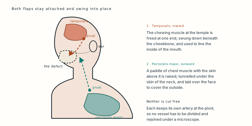
The important property is what both flaps share. Neither was detached.
A free flap is cut completely away from the body, carried to the defect, and its artery and vein are sewn to new vessels there under a microscope. It is what allowed a jaw to be rebuilt from a leg in another case here. It also requires a microscope, instruments for vessels a couple of millimetres across, a surgeon trained in it, and hours of theatre time.
A pedicled flap never leaves its own blood supply. It stays joined at one point, the pedicle, and rotates around it like a door on a hinge. The limit is reach: the tissue can only travel as far as the arc of that rotation allows, which is why the anatomy of each flap determines which defects it can be used for.
What that buys is independence from equipment. Two pedicled flaps rebuilt a midface in a hospital where free tissue transfer was not on the table.
The test of whether it worked
The measure of a reconstruction like this is not how it looks. It is whether the mouth works.
By the time the visiting team left, he was eating a regular diet, which is the outcome that matters: the lining had sealed, the mouth held, and the flap was surviving where it had been placed. On later follow-up the temporalis flap had epithelialised, meaning the muscle surface inside his mouth had grown a lining of its own and begun to behave like the tissue it replaced.
That is the quiet part of flap surgery. Muscle moved into the mouth does not stay muscle. Over weeks it takes on a surface appropriate to where it now lives.
What the pathology meant
The tumour was an epithelial-myoepithelial carcinoma, an uncommon salivary gland cancer that takes its name from the two cell types that make it up, mirroring the two cell layers of the normal salivary duct.
It is usually low grade and it is not the most aggressive thing that can arise in a parotid. What it does do is recur locally, which is why the plan included radiotherapy despite the resection having been radical. Radiation here is not treating disease anyone can see. It is treating the possibility of cells left behind at a margin.
That distinction matters for a patient who feels well and is eating normally, because the argument for going through radiotherapy is entirely about a risk rather than a symptom.
What this case teaches
The parotid gland has the facial nerve running through it, so the first decision in any tumour there is whether the nerve survives, and a mass that has grown into the mouth answers that question for you. What follows is a defect with two faces, an outside that needs covering and an inside that needs a lining capable of living against saliva. Both were rebuilt with muscle that was never disconnected: the temporalis swung down into the mouth, the pectoralis major tunnelled up to the cheek, each rotating around its own artery. No microscope, no vessels rejoined, and a man eating a normal diet within days. Free tissue transfer is the more versatile tool and it is not the only one, which matters in most of the world.
Written from a first-person account published by the operating surgeon, Brian Camazine, MD, of the Earthwide Surgical Foundation, describing a patient treated in September 2013 and his later follow-up. It is a surgeon's own account rather than a peer-reviewed case report: no imaging, operative detail or long-term outcome is recorded beyond what is quoted. The anatomy of the parotid and facial nerve, the behaviour of epithelial-myoepithelial carcinoma and the technique of temporalis and pectoralis major flaps are drawn from standard references. The clinical photographs accompanying that account are held by the foundation and are not reproduced here. The diagram is original to MedicaseHub and may be reused freely. This article is not medical advice. Read the full disclaimer.
Related cases
- He was a face transplant candidateJaws rebuilt from a leg, a nose from a forehead, and lips that decided everything.
- The surgery was free. The cure was not.A visiting team removed a childhood sarcoma at no cost. The chemotherapy that decides whether she lives was months away.
- It was never cancer. It still had to come out.Benign and harmless are different words when the skull has no spare room.