Purple ears: the adulterant, not the drug
A painful rash spread across her face and ears over two days. Her neutrophils were falling. Her antibody panel came back pointing in two directions at once, which is not how autoimmune vasculitis usually behaves. The pattern named the cause, and the cause was not the cocaine.
- Specialty
- Dermatology, toxicology, rheumatology
- Patient
- Woman, 54
- Presentation
- Painful spreading rash over 2 days: face, ears, breasts, limbs
- Neutrophils
- 1070 per cubic mm (low)
- Lymphocytes
- 680 per cubic mm (low)
- ANCA
- Positive in both perinuclear and cytoplasmic patterns
- Anti-PR3
- Titre 1:320
- Cause
- Levamisole, a cocaine adulterant
- the cutlevamisole in cocaine
- day 0–2rash spreads
- bloodsantibodies point two ways
- stop itself-limiting
What levamisole is doing in cocaine
Levamisole was developed as a veterinary and human antiparasitic drug, and was later used briefly in cancer treatment. It was withdrawn from human use in North America because it caused agranulocytosis, a collapse of the white cells that fight bacterial infection.
It then reappeared in the drug supply. It is a cheap white powder that mixes invisibly into cocaine, but it is not simply bulk filler: it appears to act on the same reward pathways and is thought to prolong the effect, which is why traffickers add it rather than something inert. At the height of the problem, surveillance suggested a large majority of cocaine seized in the United States contained it.
So the person using the drug has no way of knowing they are also taking a compound that can destroy their skin and their bone marrow.
Why the rash has that shape
The word used for it is retiform, meaning net-like. What appears is a branching, angular, map-like pattern with jagged borders, quite unlike a general redness or a scatter of spots, and the shape is diagnostic in itself.
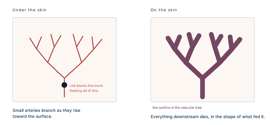
The reason is that the pattern is a picture of the blood vessels underneath. Small arteries branch as they rise toward the skin surface, each supplying its own downstream territory. When clots block those vessels, everything downstream dies, and the dead area takes the exact branching shape of the tree that fed it.
Why the ears
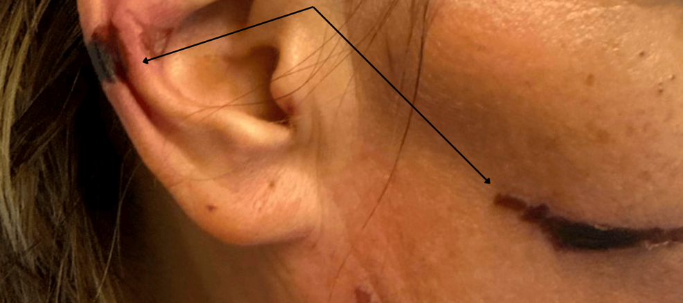
Ear involvement is the single most characteristic feature, and in published series it is the finding that most reliably points to levamisole rather than to a primary vasculitis.
The ear is an unforgiving piece of anatomy for this. It is cold, it sits at the end of the circulation, it is thin skin over cartilage with very little tissue to buffer an interruption, and its blood supply has few alternative routes. Anything that blocks small vessels shows up there first and worst. In severe cases the outer ear becomes frankly necrotic.
The antibody result that gives it away
This is the part of the case worth understanding properly, because it is where the diagnosis is usually made or missed.
ANCA testing by immunofluorescence produces one of two patterns, perinuclear or cytoplasmic. In genuine ANCA-associated vasculitis, patients are typically positive for one. This patient was positive for both, with an anti-proteinase-3 titre of 1:320.
Dual positivity is unusual in primary autoimmune vasculitis and common in levamisole-induced disease, where patients also frequently show antinuclear antibodies and antiphospholipid antibodies. A panel that lights up in every direction at once is less a sign of severe autoimmunity than a sign that something is driving the immune system indiscriminately.
Set beside purpura on the ears and a falling neutrophil count, the picture is close to specific.
Why the diagnosis matters more than it looks
Mistaking this for granulomatosis with polyangiitis has real consequences. That diagnosis leads to long-term immunosuppression, which in a patient whose neutrophils are already being destroyed is the wrong direction, and it leaves the actual cause in place.
The neutropenia is the part that can kill. Levamisole can drive the neutrophil count down to levels where ordinary bacteria become life-threatening, and the skin lesions are often the warning that arrives first.
Treatment
The central treatment is stopping exposure. Levamisole clears quickly, and once it is gone the process is self-limiting: the neutrophil count generally recovers over days to weeks, and the skin lesions settle. Reported cases have used supportive care, with steroids and, where the marrow is slow to recover, granulocyte colony-stimulating factor.
What does not resolve is tissue that has already died. Full-thickness necrosis, particularly of the ears, may need debridement and reconstruction, and severe cases have progressed to secondary infection and limb loss.
Recurrence follows re-exposure. That makes this a diagnosis where talking about where the drug came from is the treatment, not a preamble to it.
What this case teaches
Three findings that individually mean little are close to diagnostic together: retiform purpura with the ears involved, neutropenia, and an ANCA panel positive in both patterns. The shape of the rash is doing real diagnostic work, because it maps the occluded vessels beneath it rather than describing a colour. And the exposure history is not a social detail to be taken at the end of the consultation: here it is the whole diagnosis, and asking for it is the intervention.
Written from the published literature on levamisole-induced vasculopathy, including case series describing ear purpura, neutropenia and dual ANCA positivity in cocaine users. Photographs from the source case are not reproduced here. The ear photograph is from a separate patient with the same diagnosis, reproduced from Olash B, “Levamisole-Adulterated Cocaine: A Case of Vasculitis and Severe Neutropenia,” Cureus 2025;17(12):e100358, under its CC BY 4.0 licence. The diagram is original to MedicaseHub and may be reused freely. This article is not medical advice. Anyone with a painful spreading rash, particularly with fever or feeling unwell, needs urgent medical assessment. Read the full disclaimer.
One case, written up properly, when there is one worth sending
No schedule and no filler. When a case turns out to have something in it that is worth the time to explain, it goes out. Unsubscribe in one click.
Free · no spam · your address is never shared · built with KitRelated cases
- Viper bite coagulopathy: the scan that showed nothingThree hours after the bite his blood could no longer clot. A normal ultrasound is what kept him out of theatre.
- Retinoblastoma: the cancer a photo can catchOver 95% survive when it is found early, around 40% when it is not. The tumour is the same.
- Wooden foreign body on CT: the scan said gasWood reads as air on a scan. The clue that overturned the report was an infection that would not settle.