MedicaseHub
The collection

33 diagrams, free to reuse

Every case on this site needed a picture that did not exist, so it was drawn. Each one explains a single mechanism: why a scar crosses a wound edge, why a drug in the blood cannot reach dead tissue, why a replanted flap drowns in blood it cannot drain.

How to use these

They are free for anyone to use, including commercially, in teaching material, presentations, articles, videos and textbooks. You do not need to ask.

The one condition is attribution: credit MedicaseHub and link back to the page you took it from. That is the whole licence, and it is CC BY 4.0.

Each is an SVG, so it stays sharp at any size and can be recoloured or edited in Illustrator, Inkscape or Figma. Use Open the SVG below any diagram to get the file itself.

If you adapt one and it would be useful to others, tell us and we will link to your version.
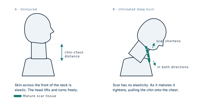
Diagram comparing an uninjured neck, where the chin clears the chest, with a burn scar contracture, where shortened scar tissue has pulled the chin down onto the chest

How contracture forms

Contracture across the front of the neck. The scar band shortens as it matures and draws the chin toward the chest, taking the lower lip and eyelids with it.
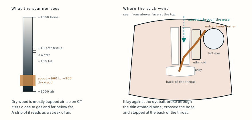
Left, a CT density scale from air to bone, with dry wood sitting close to air and well below fat. Right, a top-down view of the head showing the bamboo entering at the inner corner of the left eye, running past the eyeball, through the ethmoid sinus and across the nasal cavity to the back of the throat, and the route it was removed by, through the nose.

Why wood reads as gas

Dry wood is mostly trapped air, which is why the first report called it gas. The path on the right is why pulling it back out through the eye was ruled out.
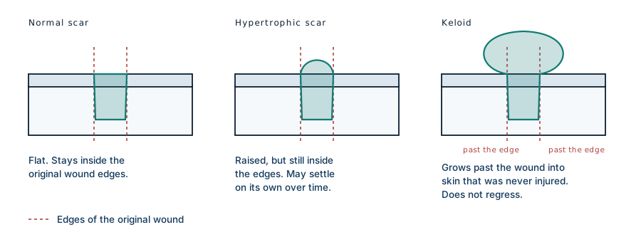
Cross-section diagram comparing three outcomes of wound healing: a normal scar sitting flat within the wound edges, a hypertrophic scar raised but still within the edges, and a keloid extending beyond the edges into uninjured skin

Scar types compared

The dashed lines mark the edges of the original wound. What separates a keloid from a hypertrophic scar is not how raised it is, but whether it crosses those lines.
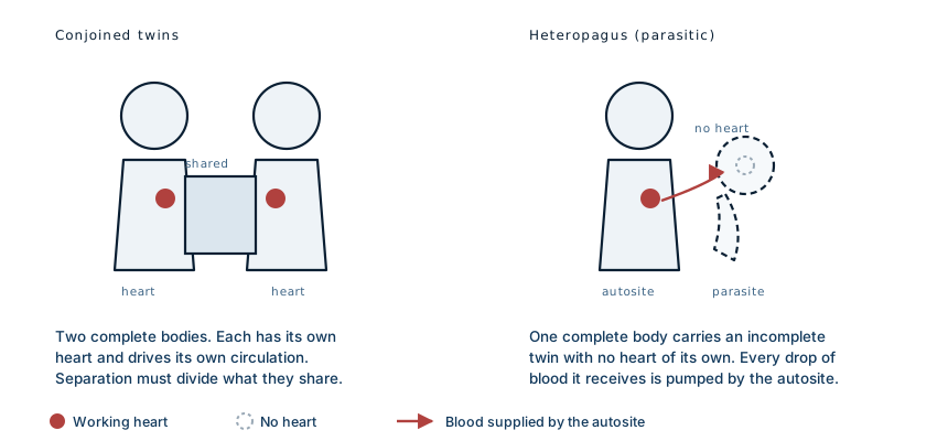
Schematic comparison. Left: conjoined twins, two complete bodies each with its own heart, joined at a shared region. Right: heteropagus twinning, one complete body with a heart carrying an incomplete twin that has no heart and is supplied entirely by the complete twin's circulation

Conjoined vs parasitic

The distinction that decides everything: in conjoined twinning there are two circulations, in heteropagus twinning there is one. The parasite has no pump and is entirely downstream of the autosite's heart.
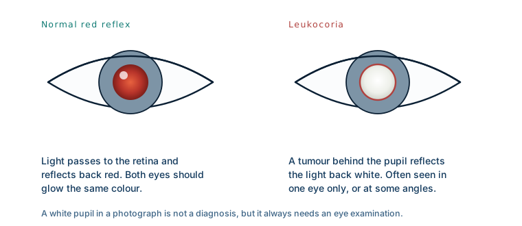
Two eyes under a camera flash compared. The left shows a normal red pupil reflex. The right shows leukocoria, a white pupil reflex, which can be the first sign of retinoblastoma

Red reflex vs leukocoria

Leukocoria often appears in one eye only, and sometimes only at certain angles, which is why it is easy to dismiss as a camera artefact.
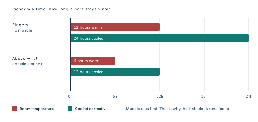
Chart comparing viability windows: fingers tolerate about 12 hours at room temperature and 24 hours cooled, while amputations above the wrist tolerate about 6 hours warm and 12 hours cooled

Ischaemia windows

Approximate working figures. Muscle is what shortens the window, which is why a forearm is a far more urgent problem than a fingertip.
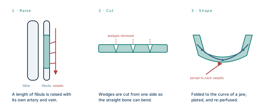
Three-stage diagram of a free fibula flap: a segment of the fibula is raised with its artery and vein, wedges are cut from one side, and the bone is folded to the curve of a jaw, plated, and its vessels joined to vessels in the neck

Free fibula flap

The free fibula flap. Norris had this done twice, once for the lower jaw and once for the upper.
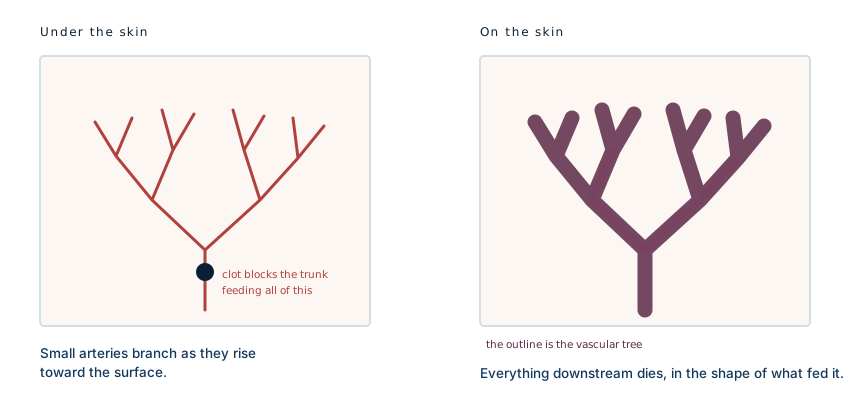
Two panels. Left: the branching network of small arteries supplying an area of skin, with a clot blocking one trunk. Right: the skin above, where the purple discolouration takes the same branching shape, because everything downstream of the clot has lost its supply

Why it is net-shaped

Retiform purpura is not a rash in the ordinary sense. It is the outline of an occluded vascular tree, seen through the skin.
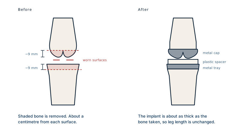
Two front views of a knee. Left: dashed lines mark cuts removing about nine millimetres from the end of the femur and about nine millimetres from the top of the tibia. Right: metal caps over both cut surfaces with a plastic spacer between them

What is removed

The implant is roughly as thick as the bone it replaces, which is why the leg does not end up shorter. Simplified: the real femoral cuts include angled chamfers that the front view cannot show.
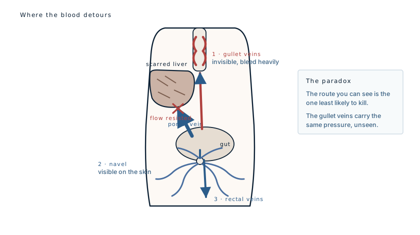
Diagram of the trunk showing blood from the gut meeting resistance at a scarred liver and detouring by three routes: upward to veins at the lower gullet, forward through a reopened vein at the navel onto the skin, and downward to veins around the rectum

Three escape routes

The three main escape routes. Only one of them shows on the outside of the patient.
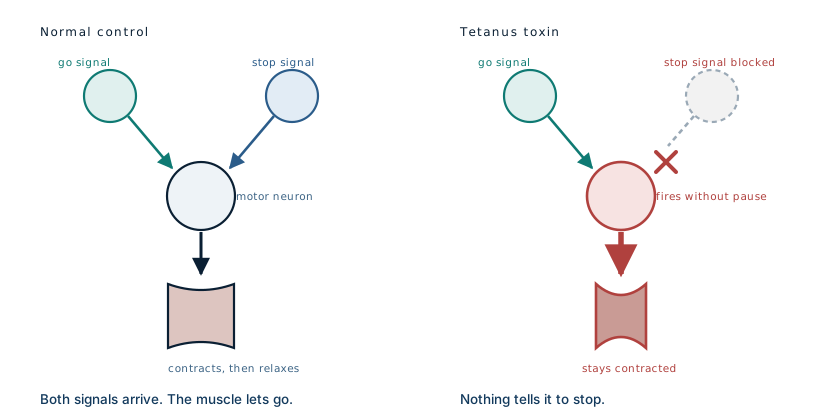
Two panels. Normal control: a motor neuron receives both a go signal and a stop signal, so the muscle contracts and then relaxes. Tetanus: the toxin blocks the stop signal, so the motor neuron fires continuously and the muscle stays contracted

How the toxin works

The toxin does not stimulate muscles. It removes the ability to stop stimulating them.
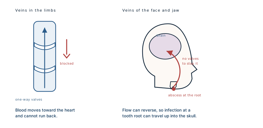
Two panels. Left: a vein in the limbs contains one-way valves, so blood cannot flow backwards. Right: the veins of the face and jaw have no valves, so infection at a tooth root can travel backwards up into the venous spaces inside the skull

Valveless veins

Most veins have valves that permit flow in one direction only. The veins draining the face and jaw do not.
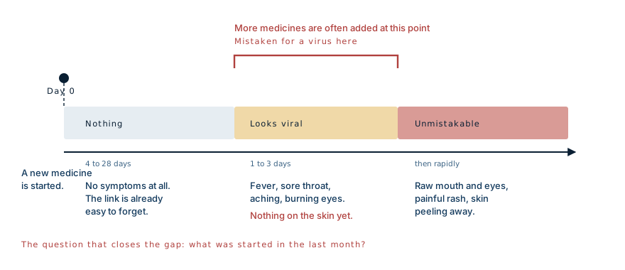
Timeline. A new medicine is started, then nothing happens for four days to four weeks. Then fever, sore throat and burning eyes begin, which is indistinguishable from a virus and where the diagnosis is missed. Only afterwards do raw mouth and eye lesions and peeling skin appear

The latency window

The gap between the drug and the symptoms is the whole problem. By the time anything appears, the medicine that caused it was started weeks ago.
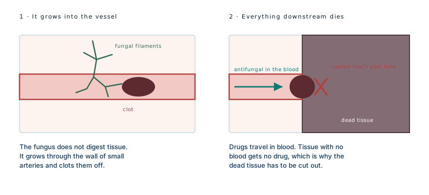
Two panels. Left: fungal filaments grow through the wall of a small artery into the channel inside it, where a clot forms. Right: the vessel is blocked, all tissue downstream has died, and an antifungal drug travelling in the blood cannot reach past the blockage

Angioinvasion

The tissue is not consumed. It is cut off. The same principle produces the net-shaped skin lesions seen in other vessel-blocking diseases.
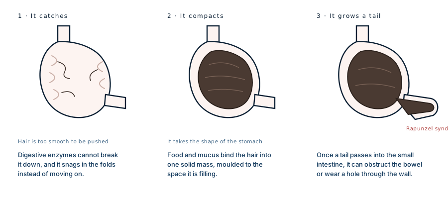
Three stages inside a stomach: individual hairs catching in the folds of the lining, then a compacted mass filling the stomach and taking its shape, then a tail of the mass extending through the stomach outlet into the small intestine

How it builds

The mass is a cast of the stomach. When a tail passes through the outlet into the small bowel, the condition is called Rapunzel syndrome.
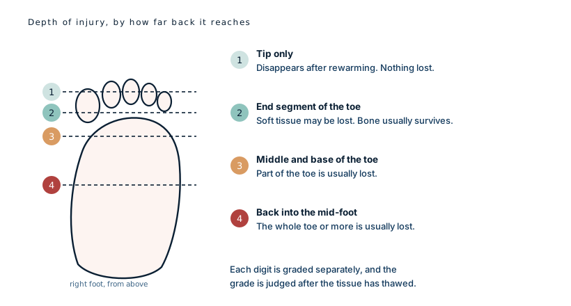
A foot seen from above with four levels marked. Grade one at the tip resolves after rewarming. Grade two reaches the end segment of the toe. Grade three reaches the middle and base of the toe. Grade four reaches back into the bones of the mid-foot

Cauchy grading

Read it back-to-front: the grade is how far the injury reaches, not how bad it looks.The Cauchy grading system. The grade predicts what will be lost, which is why it is recorded digit by digit.
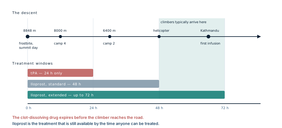
Timeline in hours from injury. Clot-dissolving tPA is effective within about 24 hours, iloprost within about 48 hours with this study extending toward 72. Climbers evacuated from Everest reach Kathmandu between 48 and 72 hours, outside the tPA window entirely

Treatment windows

The two bars are drug windows. The block on the right is when a climber can physically arrive.His arrival at 66 hours sits in the zone this study was testing. The treatment with the strongest evidence had expired more than two days earlier.
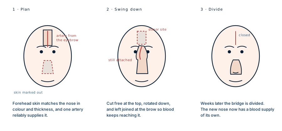
Three stages of a paramedian forehead flap. A strip of forehead skin is outlined over the artery running up from the inner eyebrow, then cut free at the top and swung down over the nose while remaining attached at the brow, and finally the connecting bridge is divided once the new nose has its own blood supply

The forehead flap

The paramedian forehead flap. Described in India well over two thousand years ago and still the standard reconstruction for a missing nose.
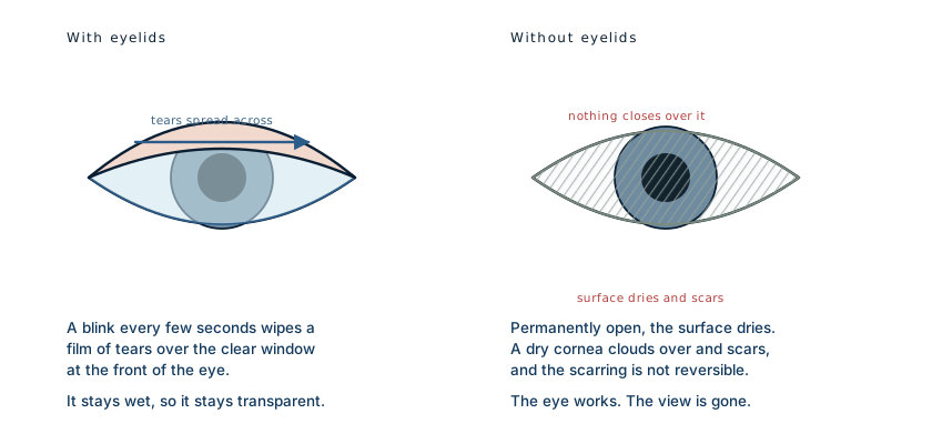
Two eyes compared. With eyelids, a blink spreads tears across the clear front surface, which stays transparent. Without eyelids the surface is permanently exposed, dries, and becomes cloudy and scarred

Why eyelids matter

The eye itself can be entirely healthy and the person can still go blind, because the surface in front of it has no protection.
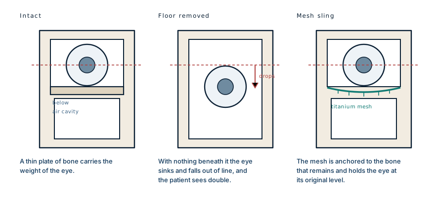
Three cross-sections through the eye socket. First the eyeball rests on a thin plate of bone above an air cavity. Second, with the plate removed the eyeball sinks and drops out of line. Third, a titanium mesh sling anchored to the remaining bone holds the eye at its original level

The orbital floor

The plate is thin enough to be nearly transparent on a scan, and it is the only thing holding the eye at the right height.
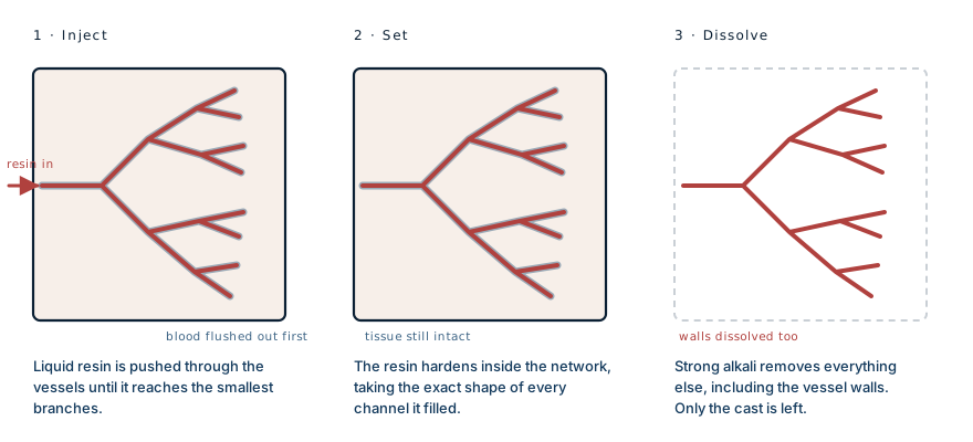
Three stages. Resin is pushed into the vessels of a specimen, it hardens inside the whole branching network, and then the surrounding tissue is dissolved away in strong alkali, including the vessel walls, leaving only the solid cast

Corrosion casting

The alkali does not distinguish between the tissue and the vessels running through it. Everything organic goes.
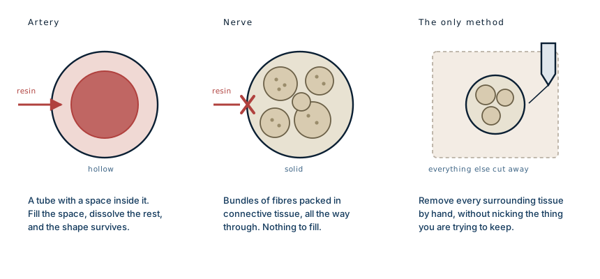
Three cross-sections. An artery is a tube with a hollow channel that resin can fill. A nerve is solid throughout, packed with bundles of fibres, so there is nothing to inject. The only method is to cut away every surrounding tissue by hand

Nerve vs vessel

A vessel is a container. A nerve is a cable. One can be filled; the other can only be uncovered.
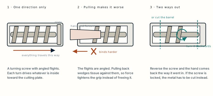
Three cut-away views of a grinder barrel. The screw drives everything toward the cutting plate. A hand pulled backwards binds against the angled flights. The only ways out are reversing the screw or cutting the barrel apart.

Why it will not come back

The flights are angled, so force applied backwards tightens the grip rather than releasing it.
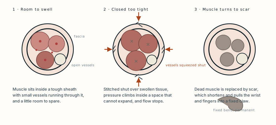
Three cross-sections of a forearm. Normally muscle sits in a sheath with open vessels and room to swell. Closed under tension, pressure rises and the vessels are squeezed shut. The dead muscle then scars and pulls the wrist and fingers into a fixed bend.

Closure under tension

The compartment cannot expand. Once the pressure inside it exceeds what the small vessels can push against, the muscle stops being perfused while everything around it still looks intact.
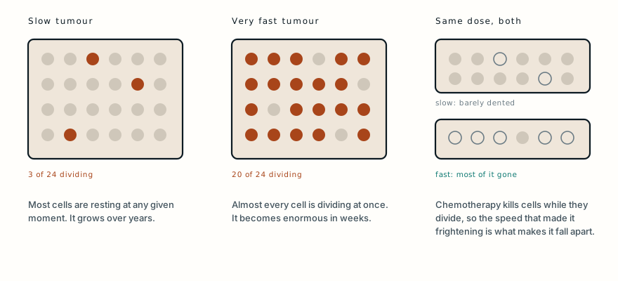
Three panels. In a slow tumour only a few cells divide at any moment. In a very fast tumour almost all of them do. Because chemotherapy kills dividing cells, the same dose barely dents the slow tumour and removes most of the fast one.

Speed cuts both ways

Chemotherapy acts on cells while they divide. A tumour in which nearly every cell is dividing is the most vulnerable target there is.
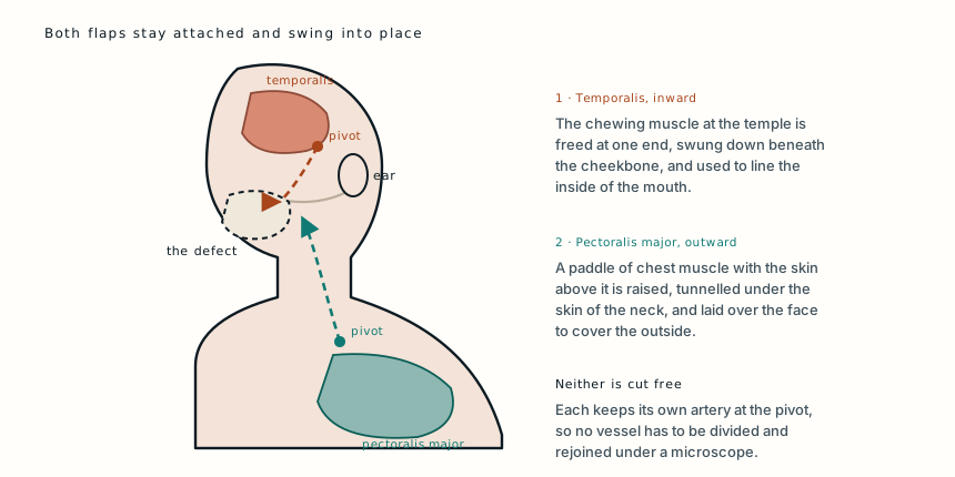
A side view of the head and chest. The temporalis muscle at the temple swings down under the cheekbone to line the inside of the mouth. The pectoralis major with its overlying skin is tunnelled up under the neck to cover the outside of the face. Each stays joined to its own artery at a pivot point.

Two pedicled flaps

A pedicled flap rotates around the vessel that feeds it. The arc of that rotation is what decides whether the tissue can reach the defect at all.
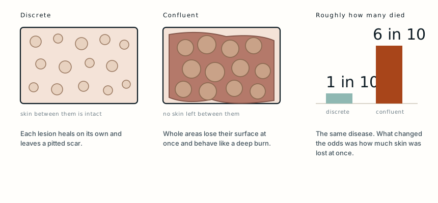
Two patches of skin compared. In discrete smallpox the pustules sit apart with normal skin between them and about one patient in ten died. In confluent smallpox they merge, destroying the skin between them, and around six in ten died.

Discrete against confluent

The classification was not descriptive. It was the closest thing the period had to a prognosis.
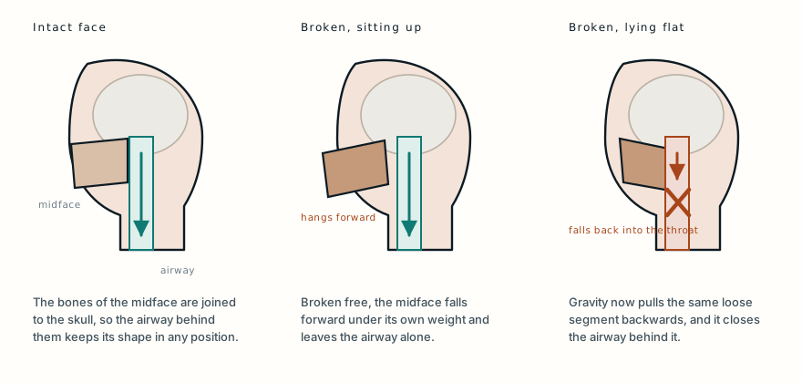
Three side views of a head. With the facial skeleton intact the airway stays open in any position. With the midface broken free it hangs forward when the patient sits up, leaving the airway clear. Lying the same patient flat lets the loose segment fall backwards and close the airway.

Why position mattered

Nothing was blocking his throat. The structure that normally holds it open had come away from the skull, and only his position was keeping it clear.
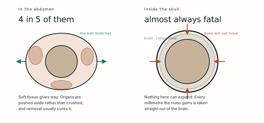
Two cross-sections. In the abdomen, where four in five of these occur, the wall stretches and organs are pushed aside. Inside the skull nothing can expand, so the mass takes its space directly from the brain.

Why the site decides it

The same growth, in two places. One is an operation with a good outcome and the other is a decade of pressure on a developing brain.
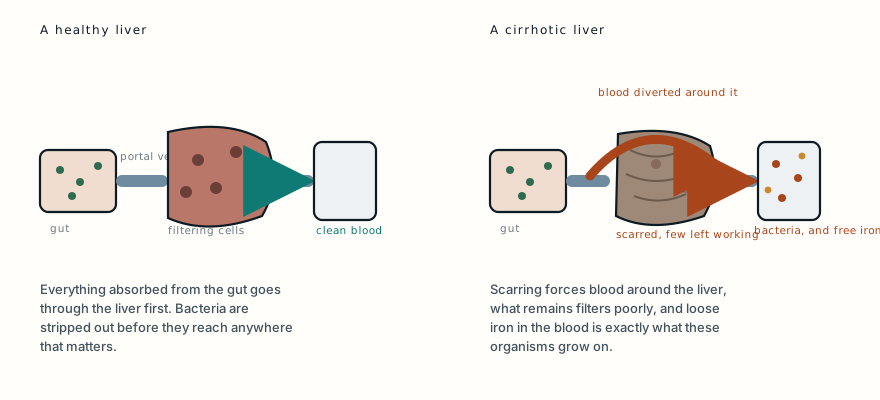
Two diagrams of the route from gut to body. Normally blood from the intestine passes through the liver, where resident immune cells strip out bacteria. In cirrhosis the blood is diverted around the scarred liver, the remaining filtering cells work poorly, and free iron in the blood feeds the organisms that get through.

Why the liver decides it

Everyone at that table swallowed the same organisms. Only one of them had lost the filter that normally deals with them.
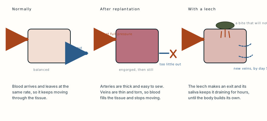
Three panels. Normally blood arrives through an artery and leaves through veins at the same rate. After replantation the artery is repaired but the thin veins are not, so blood fills the tissue and stops moving. A leech makes an exit whose bite keeps oozing for hours, draining the tissue until new veins grow.

In, but not out

The flap was not short of blood. It was full of blood that had no way out.
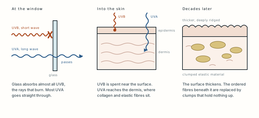
Three panels. Window glass blocks almost all ultraviolet B, the short wavelength that burns, but lets most ultraviolet A through. In the skin, ultraviolet B is spent near the surface while ultraviolet A reaches the dermis where collagen and elastic fibres sit. Decades later the surface has thickened and the fibres beneath it have been replaced by clumps of damaged elastic material.

Through the glass

The glass removed the warning, not the exposure. What got through went deepest.
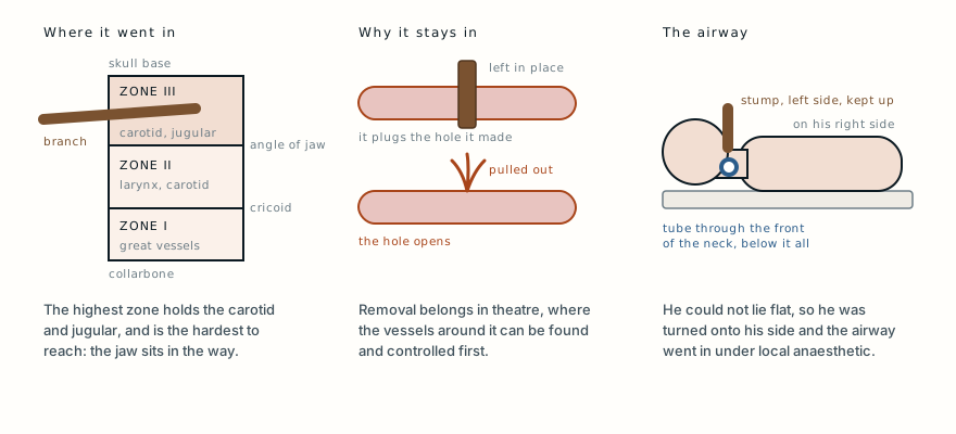
Three panels. The neck is divided into three zones, and the branch entered the highest one, between the angle of the jaw and the skull base, where the carotid artery and jugular vein run. An object left in place plugs the hole it has made in a vessel; pulled out, the hole opens. Because the stump on the left side of his neck stopped him lying flat, he was turned onto his right side and the airway was placed through the front of the neck.

Left in, on his side

The branch sat in the part of the neck that is hardest to reach, and it stayed in until someone could reach it.

One case, written up properly, when there is one worth sending

No schedule and no filler. When a case turns out to have something in it that is worth the time to explain, it goes out. Unsubscribe in one click.

Free · no spam · your address is never shared · built with Kit