The tumour covered half her face. Surgeons rebuilt it from the tumour’s own skin.
A 74-year-old woman arrived with a tumour 13 centimetres across that had swallowed the right side of her face, covering her eye, her cheek and the side of her nose. It was the largest of its kind ever recorded. Her surgeons cut off its blood supply, removed it in one piece, and closed her face with the skin that had stretched over it.
- Specialty
- Plastic and maxillofacial surgery
- Patient
- Woman, 74, with untreated high blood pressure and atrial fibrillation
- Tumour
- Myoepithelial carcinoma of the ethmoid sinus, low grade
- Size
- 11.5 × 8.8 × 13.3 cm, the largest of its type on record
- Effects
- Loss of vision on the right, blocked nose, upper jaw destroyed
- Before surgery
- Feeding arteries blocked, 2 days earlier
- Operation
- Whole tumour removed; face closed with its own preserved skin, in one stage
- Follow-up
- 3 years, no recurrence
- Left with
- A lower eyelid turned outwards, not corrected because of the cost
- arrivalhalf a face
- day −2the blood supply
- surgeryout in one piece
- 3 yearsno recurrence
Thirteen centimetres
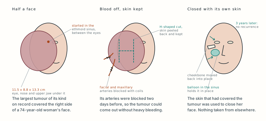
In 2019 a 74-year-old woman was admitted to the Vietnam National Burns Hospital in Hanoi with a tumour on her face. It measured 11.5 by 8.8 by 13.3 centimetres and occupied nearly half of the right side of her face. It had grown over her right eye, pressing on it until she had lost the sight on that side, and pushed against her nose until it blocked it. The skin stretched over it had a good blood supply and was not inflamed or broken.
She also had high blood pressure and atrial fibrillation, an irregular heart rhythm, and neither had been treated. That made any long operation with heavy bleeding much more dangerous for her.
A CT scan showed where the tumour had started: in the ethmoid sinus, a honeycomb of small air cells between the eyes, at the root of the nose. From there it had grown outwards into her right cheek and invaded her right maxilla, the bone of the upper jaw, destroying it.
A biopsy identified it as a myoepithelial carcinoma. Myoepithelial cells are contractile cells that sit around the small ducts of glands, and tumours made of them usually arise in the salivary glands. Only about 100 cases of myoepithelial carcinoma had been reported anywhere by early 2022, and only 14 in the nose and sinuses, none of them larger than 8 centimetres. This one was low grade, with very few dividing cells, which usually means slower growth and a low chance of spread. It was simply enormous.
Shutting off the supply first
A tumour this size is fed by a dense network of blood vessels, and cutting into it can cause torrential bleeding. For a 74-year-old with an untreated heart rhythm problem, that was a real threat.
So before operating, the team embolised it. Working through the blood vessels from inside, they blocked the right facial artery and the right maxillary artery, the main vessels feeding the tumour, with pieces of gelatin sponge and metal coils. The aim was to reduce blood loss and shorten the operation, and to give them time to bring her heart conditions under control. Two days later her heart was stable, and she was ready for surgery.
An H on the skin
The tumour turned out to be encapsulated, wrapped in its own capsule with clear borders, which made it possible to remove completely. The surgeons began with an H-shaped incision through the skin over the middle of the tumour, and dissected the skin back in four directions. They were careful to keep as much of that skin as they could, because they would need it later.
Once the tumour was exposed, they tied off the facial and maxillary vessels they met, controlled the smaller bleeding points, and removed every part of the tumour along with the dead tissue it had left in the right eye socket, the nose, the cheek and the maxillary sinus. They tried to preserve the right eyeball itself.
Then came the rebuilding. Removing a tumour this size leaves a large hollow, and the skin needs something to lie on. The surgeons used her own right cheekbone as the framework: with a hooked instrument they lifted, realigned and reshaped it. To hold it in place, they slid a Foley catheter, a tube with a small balloon on its tip, into the maxillary sinus and inflated the balloon, so that it pushed the bone and the tissues over it out into position and also let fluid drain.
They sealed off the back opening of the right nasal passage, which had lost its function, with a flap of lining from inside the nose. They removed the surface layer from part of the local skin so it could be tucked in as padding. And then they brought the skin that had covered the tumour forward over the defect as an advancement flap, rebuilding the lower eyelid and lips. No tissue had to be taken from anywhere else on her body. The whole thing was done in a single operation.
Three years later
Because the tumour had been removed completely, was low grade and had had its blood supply cut, the team decided she did not need radiotherapy afterwards.
Three years after the operation she had had no complications and no recurrence, and the authors describe the cosmetic result as satisfactory. Her face had its shape back. She was left with ectropion of the lower eyelid, the lid turning outwards away from the eye, which is a common result when the tissue below the eye has been stretched or rebuilt. It could have been corrected with another operation, but she declined because of the cost.
The photographs of her before the operation and three years after are in the original paper, as Figures 1 and 5. They are not reproduced here because of the paper’s licence, but they are worth looking at.
Why such a big tumour could come out so cleanly
The size of a tumour is not the same as how dangerous it is. This one had grown slowly, stayed inside its own capsule and not spread, so once its blood supply was dealt with, it could be lifted out whole. Many smaller tumours of more aggressive types are far harder to remove.
Myoepithelial carcinomas in the sinuses have been removed both through open surgery and endoscopically, through the nose, but a tumour this large and this far outside the sinus needed an open operation. The authors note that whether to give radiotherapy afterwards is still debated, and is usually reserved for tumours at higher risk of spreading. The longest follow-up they found in the literature was 40 months without relapse, in a patient whose tumour was only 3 centimetres across.
The other lesson is in the skin. A slow-growing tumour stretches the skin over it, and that stretched skin is exactly the extra tissue a surgeon needs to close the hole it leaves. Keeping it, rather than cutting it away with the tumour, let her face be rebuilt in one operation without borrowing tissue from anywhere else.
What this case teaches
A 74-year-old woman with untreated high blood pressure and atrial fibrillation arrived with a low-grade myoepithelial carcinoma of the ethmoid sinus, 11.5 by 8.8 by 13.3 centimetres, the largest of its type on record, covering the right half of her face and costing her the sight in her right eye. Her surgeons blocked its main feeding arteries two days before surgery, opened the skin over it in an H and kept that skin, removed the encapsulated tumour completely, reset her cheekbone and held it with a balloon in the sinus, and closed her face with the tumour’s own skin in a single operation. Three years later there was no recurrence, and her only lasting problem was a lower eyelid turned outwards that she chose not to have fixed. A tumour that has grown large slowly can sometimes leave behind the very tissue needed to repair the damage it has done.
Was this case worth your time? Buy the next one a coffee ($3), or support MedicaseHub monthly.
Next case · MC-048 He lived with it for 20 years. It weighed 20 kilograms.A 90-year-old man refused treatment for a slow-growing lump on his hip for more than 20 years. When it began to affect his walking, surgeons removed a benign fatty tumour of 20 kilograms.Read it →
Want the next case like this?
When a new case is written up, it comes to your inbox: the story, the diagram and what it teaches. No schedule and no filler. Unsubscribe in one click.
Free · no spam · your address is never shared · built with KitRead the original paper: doi.org/10.7181/acfs.2023.00374
Adapted from Vu QV, Hoang TT, Tran VA, Tong TH and Nguyen HH, “Successful surgical interventions for a giant and complicated myoepithelial carcinoma: a case report,” Archives of Craniofacial Surgery 2024;25(4):197–200, from the Vietnam National Burns Hospital and Viet Duc Hospital, Hanoi. The presentation, scan and biopsy findings, the embolisation, the operation and reconstruction, and the three-year outcome are as reported there, summarised in the author’s own words; the figures on how rare the tumour is and the points on radiotherapy are those the authors give, and the explanations of the anatomy and procedures are from standard references. The authors report that the patient gave written consent to publication. The paper is published under a Creative Commons Attribution Non-Commercial licence (CC BY-NC 4.0), so its photographs are not reproduced here; they can be seen in the original paper. The diagram is original to MedicaseHub, is simplified, and may be reused freely under CC BY 4.0. This article is not medical advice. Read the full disclaimer.
Related cases
- He lived with it for 20 years. It weighed 20 kilograms.Another tumour that grew huge slowly, and came out whole.
- Half his face was torn away. It was all put back in one operation.Another half of a face rebuilt in a single operation.
- It was never cancer. It still had to come out.Another growth in the head where size, not malignancy, was the problem.