Half his face was torn away. It was all put back in one operation.
A 21-year-old motorcyclist was hit by a fast-moving car. The left side of his face, from the forehead to the chin, was torn and peeled back, and blood and loose tissue were blocking his airway. The team got a tube into his windpipe, and within 45 minutes of his arrival surgeons were putting every layer of his face back where it belonged.
- Specialty
- Emergency medicine, oral and maxillofacial surgery, ophthalmology
- Patient
- Man, 21, motorcyclist
- Mechanism
- Collision with a fast-moving car
- Arrival
- Six hours after the crash, bleeding, not fully alert, blood pressure 91/41, pulse 130
- Torn
- Forehead, left eyebrow and eyelids, nose, left cheek, part of the right cheek, both lips, chin
- Spared
- The left eye, its sight and its movements
- Airway
- Blocked by blood and loose tissue; tube placed after suction
- Scans
- A small epidural bleed, a crack in the left upper jaw that had not shifted, neck spine normal
- Theatre
- Within 45 minutes of arrival; one operation, layer by layer
- Home
- Fourteen days later, wounds healing without tissue loss
- 0 hthe crash
- 6 hhe reaches hospital
- airwaya tube past the damage
- scanswhat else was broken
- 45 minevery layer back
- 14 dayshome
A motorcycle and a fast car
He was 21 and riding a motorcycle when a fast-moving car hit him. The impact tore the soft tissue of his face, mostly on the left side, and peeled it away from the structures underneath.
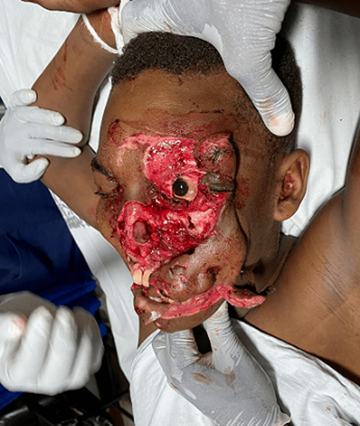
Two words describe injuries like this. Avulsion means tissue torn away from where it belongs. Degloving means skin and the tissue beneath it stripped off the deeper layers, the way a glove comes off a hand. He had both, across the forehead, the left eyebrow, both left eyelids, the nose, the whole of the left cheek, part of the right cheek, both lips and the skin over the chin.
Injuries of the face this severe are dangerous for reasons that have nothing to do with how they look. They bleed heavily, they can block the airway, and the force behind them can injure the brain and the neck.
Bleeding, not fully alert, heading into shock
He was referred to the emergency department of Muhimbili National Hospital in Dar es Salaam six hours after the crash. His wounds were still bleeding, and he was not fully alert: his Glasgow Coma Score was 13 out of 15.
His blood pressure was 91 over 41 and his pulse 130 a minute. He was breathing 28 times a minute. Low pressure with a racing heart is the body compensating for blood it has lost, and it was one of the two things that could kill him first.
The other was his airway. Blood from the wounds and loose flaps of his own tissue were blocking it. His oxygen saturation was 99% on room air, a reminder, as in another face injury on this site, that a normal oxygen reading says nothing about what the airway will do in the next few minutes.
One thing had been spared. Beneath the torn left eyelids, the left eye itself was intact, and so were its sight and its movements.
Clinical photograph · graphicHis face on arrival, before the airway was secured. This is a severe facial injury. Tap to show.
Why a mask would not work
The usual first step for a patient who cannot keep an airway open is a bag and mask, pressed over the nose and mouth to push air in. That depends on the mask sealing against the face. His face was torn and bleeding, and there was nothing for a mask to seal against.
Loose flaps of tissue add a second problem. They can fall into the airway and behave like a foreign body. Patients with injuries like his usually need a tube placed early.
The team prepared for the worst case. A tray for a cricothyrotomy, a surgical opening through the front of the neck, was ready, along with a video laryngoscope, a camera that lets the doctor see the vocal cords around an obstruction. After several rounds of suction and putting the displaced tissue back into position, they were able to pass a tube into his windpipe, using a rapid sequence intubation, the standard emergency technique of giving sedation and a muscle relaxant together.
Checking what the face was hiding
An ultrasound at the bedside, looking for blood in the chest and abdomen, was negative. The CT scan of his head and neck found a collection of blood under the scalp, a small collection of blood on the left side between the skull and the membrane that covers the brain, an epidural haematoma, and a crack in the front of the left upper jaw that had not moved out of place, with blood in the sinus behind it. His neck spine was normal, and X-rays of his pelvis and left ankle were too.
His haemoglobin was 10.4 g/dL. He was given two litres of saline and two units of whole blood, and antibiotics, ceftriaxone and metronidazole, because the wounds were heavily contaminated, exposed the cartilage of the nose and were connected to the jaw fracture. His tetanus vaccination history was unknown, so he was given a tetanus shot.
The jaw fracture needed nothing more than pain relief. A crack that has not shifted out of position usually does not need urgent repair, and his caused no problems with function or appearance.
One operation, from the inside out
Within 45 minutes of reaching the emergency department he was in the operating theatre. Under general anaesthetic the skin was cleaned with iodine and the wounds were washed out with saline and diluted iodine. Contaminated tissue was cut away, but as little as possible: every piece of his own tissue that could be saved was worth more than anything that could replace it.
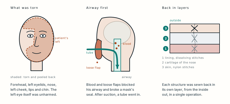
Want this diagram print-ready? It is in the diagram pack.
Then the face was rebuilt structure by structure, in layers. The deep tissues, the lining of the mouth, the lips, the chin, the corner of the mouth, the side of the nose and its base, were sewn with stitches that dissolve. The nose was rebuilt from the inside out, first its lining, then its cartilage framework, and the right side of the nostril was moved back into place before the skin over it was closed. The skin everywhere was closed with nylon.
Layering matters because a face is not one sheet. Lining, cartilage and skin each have their own job, and if they are not put back separately and in order, the result can scar, collapse or pull out of shape.
The eyelids went to an ophthalmologist. They contain the glands that make tears and the small channels that drain them, and those need to be repaired correctly, which can be done within 48 hours of the injury. A cut near the inner corner of the right eye was repaired in layers. In the left eyelids the tear-producing and lid glands were put back and stitched in layers, and the edges of the eyelids were lined up and closed. The rest of the skin was then laid back into position and sewn, the bleeding was stopped, and the eye was covered with antibiotic ointment and a pad.
A face that healed without losing tissue
He went from theatre to the surgical intensive care unit. The next day he was taken off the ventilator and the breathing tube was removed. After three uneventful days in intensive care he moved to the oral and maxillofacial ward, where he continued to receive pain relief, antibiotics, fluids and wound care.
The stitches came out nine days after the operation. The wounds healed without any tissue dying, which is the complication that flaps of torn tissue are most at risk of. An eye examination found nothing wrong with his vision. He went home fourteen days after he arrived, with follow-up in the oral and maxillofacial clinic.
Clinical photographHis face after the stitches came out. Tap to show.
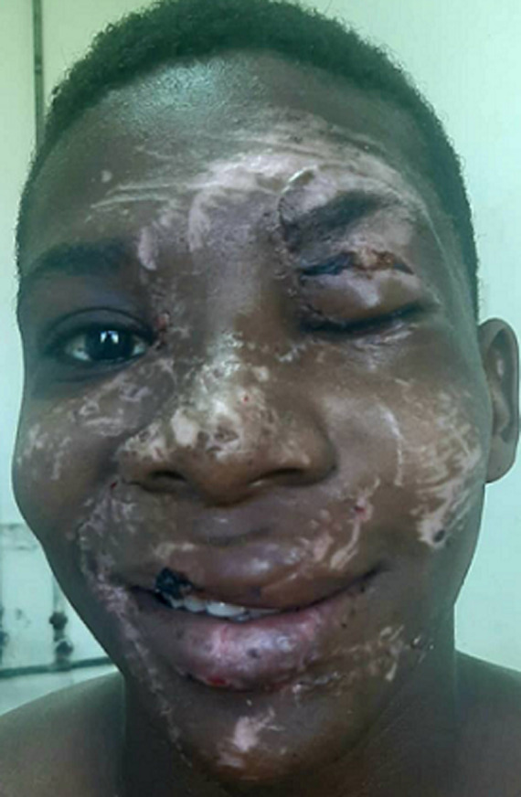
He remembered nothing of the crash. In a statement published with the case, he described asking the emergency physician, a week later, to show him the photographs taken before the repair, and feeling close to tears and full of gratitude when he saw them. “They re-created my face and gave me a second chance to live,” he said, and asked that his experience be used to teach others.
Why repair it all at once
It might seem safer to repair an injury this large in stages. The authors argue the opposite. Once the threats to life are under control, an early single-stage primary repair gives better function and a better appearance, with less infection and less scarring than delayed reconstruction. The wounds are cleaned and repaired without delay, and as much of the patient's own tissue as possible is kept.
It also takes several specialties at once. In his case the emergency physicians secured the airway and the circulation, the oral and maxillofacial surgeons rebuilt the face, and an ophthalmologist repaired the eyelids, with each consulted as soon as the first assessment showed what was involved.
The other cases on this site with devastating facial injuries ended in face transplants. The difference is what was left. When tissue has been destroyed or lost, there is nothing of the patient's own to put back. Here almost everything was still there to be repositioned, and a single operation gave him back his own face.
What this case teaches
The injury that looked worst was not the one that threatened his life first. He arrived six hours after the crash with half his face torn and peeled back, but the dangers were blood loss and an airway blocked by blood and his own loose tissue, and his oxygen reading of 99% gave no warning of either. A mask could not seal against his face, so the team prepared a surgical airway and a video laryngoscope, cleared the airway with suction, put the displaced tissue back and passed a tube. Only then did the face come next: within 45 minutes of arrival he was in theatre, where each structure was cleaned, trimmed as little as possible and sewn back in its own layer, lining first and skin last, with the eyelids repaired by an ophthalmologist. He was off the ventilator the next day and home in two weeks, with every structure back in its place.
Read the original paper: doi.org/10.11604/pamj.2022.41.158.32802
Adapted from Simbila AN, Laizer PJ, Waihenya WN, Deoglas DK, Mwashilemo AB and Kiwale DJ, “Traumatic hemifacial avulsion and degloving injury: a case report,” The Pan African Medical Journal 2022;41:158, from the Departments of Emergency Medicine, Oral and Maxillofacial Surgery and Ophthalmology at Muhimbili National Hospital and Muhimbili University of Health and Allied Sciences, Dar es Salaam, Tanzania. The history, examination, vital signs, imaging, treatment, operative steps, timeline and outcome are as reported there, summarised in the author’s own words; the patient’s quoted words are from the patient perspective published with the case. The points on airway management, single-stage repair, eyelid repair and antibiotics are drawn from the paper’s discussion, and the explanations of avulsion, degloving, the Glasgow Coma Score and intubation from standard references. The paper is published under a Creative Commons Attribution 4.0 licence, and the two clinical photographs are reproduced from it under that licence, unchanged; the authors report written consent from the patient for publication of the images. The diagram is original to MedicaseHub, is simplified, and may be reused freely under CC BY 4.0. This article is not medical advice. After a serious facial injury, call emergency services and, if the person is conscious, let them stay in whatever position they can breathe in most easily. Read the full disclaimer.
Find these cases useful? Support MedicaseHub on Patreon.
One case, written up properly, when there is one worth sending
No schedule and no filler. When a case turns out to have something in it that is worth the time to explain, it goes out. Unsubscribe in one click.
Free · no spam · your address is never shared · built with KitRelated cases
- He could breathe sitting up, but not lying downAnother face that hit something at speed, and an airway that looked safer than it was.
- He was not going blind from the fireWhat happens when facial tissue is lost rather than torn, and there is nothing of the patient’s own to put back.
- They cut the branch down. Nobody pulled it out.A branch through the face, and the same rule: the airway and the bleeding come before anything that shows.