They cut the branch down. Nobody pulled it out.
A road accident drove a wooden branch into the side of a young man's face, below the jaw, and out through his mouth. It was about eight feet long. The first hospital sawed it down to two feet and sent him on with the rest still inside him, and that was the right decision.
- Specialty
- Oral and maxillofacial surgery, emergency medicine
- Patient
- Man, 30, Kottayam, Kerala
- Mechanism
- Road traffic accident; a wooden branch entered the left side of the face
- The object
- About 8 feet long; cut to about 2 feet before transfer
- Path
- In below the angle of the left jaw, out through the mouth
- On arrival
- Conscious, short of breath, bleeding into mouth, nose and throat; BP 140/90, pulse 110
- The airway problem
- Could not lie flat, because the branch was lodged behind his neck
- Airway secured by
- Tracheostomy under sedation and local anaesthetic, lying on his right side
- Removal
- In one piece, through the broken jaw, with the facial and lingual arteries tied
- Spared
- Carotid artery, jugular vein, facial and lingual nerves
- Discharged
- Day 10, breathing tube removed
- 0 hthe branch goes in
- transfereight feet to two
- arrivalhe cannot lie down
- airwayon his side
- theatrethrough the fracture
- CTwhat wood leaves behind
- day 10home
In below the jaw, out through the mouth
He was 30, and he was hurt in a road accident. The report does not describe the crash itself, only its result: a wooden branch had gone into the left side of his face just below the angle of the jaw and come out through his mouth.
On the way through it broke his lower jaw in several places, on the left side of the body of the mandible and at the front, where the two halves meet.
The place it went in is what made this dangerous. Surgeons divide the side of the neck into three zones, and the highest of them runs from the angle of the jaw up to the base of the skull. The internal and external carotid arteries pass through it, along with the jugular vein and several of the cranial nerves that control swallowing, the tongue and the face. It is also the hardest zone to operate in, because the jaw and the skull base sit in the way of the surgeon.
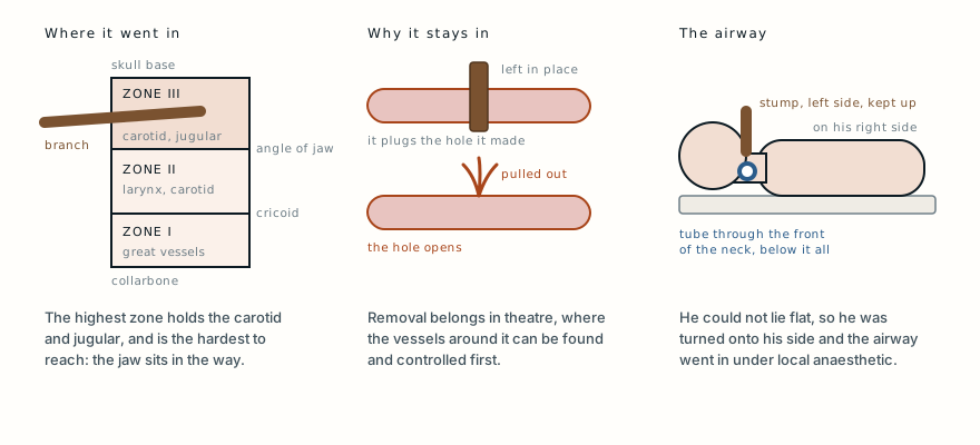
Nothing that large passes through that space by luck alone. In his case it had, so far, missed the great vessels. Nobody could know that yet.
Why the first hospital left it in
The branch was about eight feet long when he reached the local hospital. It left there about two feet long, with the rest still in his face, and he was sent on to the oral and maxillofacial surgery team at Medical College Kottayam.
Pulling it out would have been the instinctive thing to do, and it is exactly what trauma teaching says not to do.
An impaled object that has torn a blood vessel is often also the thing pressing that tear closed. While it stays where it is, bleeding is limited to what can leak around it. Removed outside an operating theatre, it leaves an open hole in a vessel that nobody can see or reach, in a part of the neck where there is no room to press on it. Removal belongs in theatre, after the vessels around the object have been found and can be controlled.
Shortening it was a separate and sensible step. Eight feet of wood cannot travel in an ambulance and cannot fit on a trolley, and every movement of the free end is transmitted, as a lever, to whatever the other end is lodged against.
Awake, bleeding into his airway
He was conscious on arrival, with no weakness or loss of sensation and no history of having been knocked out. He was short of breath and struggling to swallow. There was significant bleeding into his mouth, nose and throat, and a steady trickle from the wound where the branch had gone in. His blood pressure was 140/90 and his pulse 110.
The team worked through the standard trauma sequence, which puts the airway before everything else, and the airway was the problem.
Blood and saliva were collecting in a throat he could not clear properly, which put him at high risk of inhaling them. The usual next step, laying the patient flat to place a tube through the mouth, was ruled out. The wood lodged behind his neck meant he could not lie on his back at all, and with the branch itself coming out of his mouth there was little room to pass a tube beside it.
Readers of MC-031 will recognise the shape of this problem: a man whose injury rules out lying flat, and an airway that therefore has to be made below the damage rather than passed through it.
A tracheostomy, done awake
The surgeons shortened the branch again, this time close to the skin behind the left side of the jaw. That turned a long, awkward lever into a short stump.
They then laid him on his right side, which kept the stump on the left uppermost and clear of the trolley, and made a tracheostomy, an opening through the front of the neck into the windpipe, under sedation and local anaesthetic.
Doing it that way round matters. Putting a patient to sleep before the airway is secured takes away his own muscle tone and his own effort to breathe, and if a tube then cannot be placed, there is no way back. With him still awake and breathing for himself, the team made the airway first. Only once the tube was in the trachea did they give a general anaesthetic, through the new opening.
With the airway secure they moved on to breathing and circulation in the usual order, and a second, fuller examination found no other injuries.
Using the injury as the way in
To get the branch out safely the surgeons needed to see the whole length of it, and the vessels next to it, from the inside.
They split the lower lip in the midline and carried the incision down to where the wound began. The jaw was already broken at the front, so instead of cutting through the bone they used the fracture itself, opening the two halves of the mandible outward like a door. That approach, known as a mandibular swing, is normally made by deliberately dividing the jaw. Here the accident had done that part.
With the floor of the mouth and the space around the branch opened up, it was removed in one piece. Two named arteries had been torn along its track, the facial artery and the lingual artery, which supplies the tongue, and both were tied off. The carotid artery, the jugular vein and the major nerves of the neck turned out to be undamaged.
The wound was washed out with dilute antiseptic, the broken jaw was put back together and fixed with small stainless steel plates and screws, a drain was placed, and everything was closed in layers.
Why they looked again after it was out
After the operation he had a CT scan of the head and neck. It showed no head injury, no injury to the cervical spine, no further facial fractures, and no remaining foreign material.
The last of those is the one that matters most with wood.
Metal and glass are straightforward to find on imaging. Wood is not. It is porous and full of air when dry, so on CT it can look like a pocket of gas rather than an object, and it is easily missed on plain X-rays. It also splinters, and it carries soil, bacteria and fungi into the wound. A fragment left behind does not stay quiet: it tends to declare itself weeks or months later as an abscess or an infection that will not settle, which is what happened in MC-006.
So the surgeons checked the track for fragments directly while it was open, and confirmed with the scan once it was closed. A wound contaminated like this is also routinely covered against tetanus.
What he was left with
After the operation both the facial nerve and the lingual nerve were working, which means his facial movement and the sensation of his tongue were intact.
His recovery was uneventful. The tracheostomy tube was removed and he went home on the tenth day. At a month he had no problems with function and no major cosmetic problem.
For an injury that went through the least accessible part of the neck, that is about as good as the outcome gets.
Four centuries of learning to explore
The report sets the case against a long history. In the sixteenth century the French surgeon Ambroise Paré tied off the torn carotid artery and jugular vein of a wounded soldier, the first recorded operation for an injury of this kind.
During the Second World War, military surgeons adopted a rule that any neck wound passing through the platysma, the thin sheet of muscle under the skin, should be explored surgically, and death rates fell. A 1956 study by Fogelman and Stewart, cited in the report, found a mortality of 6% in patients explored straight away against 35% in those explored late or not at all.
Practice has become more selective since then. Stable patients with some neck wounds are now often assessed with CT angiography rather than taken straight to theatre. An object still sitting in the wound, with a patient bleeding into his airway, is not one of those cases.
What this case teaches
An object impaled in the neck is often holding shut the damage it has done, so the first rule is to leave it in: shorten it if it has to travel, but remove it only in theatre, once the vessels around it can be controlled. This branch entered the highest zone of the neck, between the jaw and the skull base, where the carotid and jugular run and access is hardest. Because the wood behind his neck stopped him lying flat and the branch filled his mouth, the airway had to be made below the injury, through the front of the neck, with him awake and on his side. The surgeons then used the jaw fracture as their route in, took the branch out whole, tied the two arteries it had torn, and scanned afterwards because wood is easy to miss and dangerous to leave behind. He went home on day ten with his face and tongue working.
Adapted from Mohan S, Varghese G, Kumar S and Subramanian DP, “Penetrating facial injury by a wooden log,” National Journal of Maxillofacial Surgery 2014;5(2):228-231, from the Departments of Oral and Maxillofacial Surgery and of Anaesthesia, Government Dental College, Kottayam, Kerala. The injury, the findings on arrival, the airway management, the operative approach and the outcome are as reported there, summarised in the author’s own words rather than reproduced. The paper gives one of the presenting complaints as “dysphasia”; in context this has been read as difficulty swallowing. The historical figures, including the Fogelman and Stewart mortality rates, are as cited in that paper. Background on the zones of the neck, impaled objects, awake tracheostomy, the mandibular swing approach and the imaging of wooden foreign bodies is drawn from standard references. The paper is published under a Creative Commons Attribution Non-Commercial Share-Alike licence, so its clinical photographs are not reproduced here. The diagram is original to MedicaseHub, is simplified, and may be reused freely. This article is not medical advice. If an object is embedded in someone, do not remove it; call emergency services. Read the full disclaimer.
One case, written up properly, when there is one worth sending
No schedule and no filler. When a case turns out to have something in it that is worth the time to explain, it goes out. Unsubscribe in one click.
Free · no spam · your address is never shared · built with KitRelated cases
- He could breathe sitting up, but not lying downAnother face injury where lying flat was impossible, and the airway went in through the neck.
- Wooden foreign body on CT: the scan said gas, it was bambooWhat happens when a piece of wood is left behind and the scan does not show it.
- The hand arrived still inside the machineAnother patient who arrived still attached to what injured him, and was freed only in theatre.