It ran from his pelvis to his neck and missed every major vessel
A 45-year-old man arrived at a trauma centre with abdominal pain. The scans showed a slender object running from his rectum up through his abdomen and chest to the right side of his neck. It was a wooden rod 63.5 centimetres long, and on its whole way through his body it had not cut a single major blood vessel.
- Specialty
- Trauma surgery, cardiothoracic surgery
- Patient
- Man, 45
- Mechanism
- Reportedly slipped in his bathtub and fell onto a broomstick
- Arrival
- Alert and stable, complaining of abdominal pain
- Object
- A wooden rod, 3 cm thick and 63.5 cm long
- Path
- Rectum, the mesentery of the bowel three times, the stomach, the diaphragm, the sac around the heart, under the right collarbone into the neck
- Blood vessels
- No vascular injury along the track
- Operation
- Chest and abdomen opened; rod cut in two and removed; 3 h 52 min
- Hospital
- Two months, with pneumonia, sepsis and weakness of the right arm
- Afterwards
- Colostomy reversed at 17 months; three operations in about 2.5 years
- 0 hthe fall
- arrivalstable, with abdominal pain
- theatrechest and abdomen opened
- 3 h 52 minout in two pieces
- 2 monthsa long recovery
- 2.5 yearsthree operations
A fall onto a broomstick
He was 45. According to the account reported, he slipped in his bathtub and fell onto a broomstick.
Impalement is a particular kind of penetrating injury, in which an object enters the body and stays there, and it typically happens when a person falls onto the object. What makes it dangerous is not only the damage along the track but what can happen when the object is moved.
A man who looked well, and a scan that did not
He arrived at a Level I trauma centre and teaching hospital alert, with a stable pulse and blood pressure. His main complaint was pain in his abdomen.
Once the trauma team had confirmed he was stable, they took X-rays and CT scans. They showed a slender object running from his rectum all the way up to the right side of his neck.
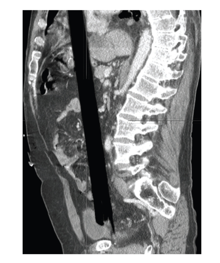
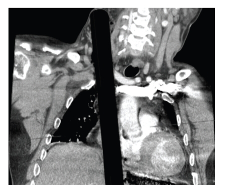
Being stable with something like this inside him was less reassuring than it sounds. An impaled object can press shut a vessel it has torn, and moving it can release that pressure. The decision was made to take him to the operating theatre to explore and treat his injuries there.
Opening both cavities before moving anything
The surgeons opened his chest through the breastbone, a median sternotomy, and his abdomen through a long incision down the middle, a midline laparotomy. Most injuries to the diaphragm are not treated through both the chest and the abdomen at once. Here it was necessary to see the full extent of the damage, and to be ready for sudden, catastrophic bleeding if moving the rod released pressure it had been keeping on the great vessels.
What they found was a blunt-ended wooden rod 3 centimetres across, with its splintered end inside the rectum. From there it had pierced the front of the rectum, passed three times through the mesentery of the small and large bowel, the sheets of tissue that carry blood vessels to the gut, gone straight through the stomach, pierced the diaphragm and the pericardium, the sac around the heart, and come to rest with its far end pinned below the right collarbone and extending into the neck.
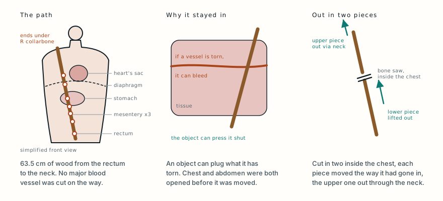
There was no injury to any blood vessel along the whole track. The authors suggest the shape of the rod explains it: its rounded end probably acted as a blunt tunneller, pushing through tissue rather than slicing it.
Cut in two inside the chest
The surgeons did not pull it back out the way it came. They cut it in two inside the chest, in the space between the lungs, with a bone saw. The lower piece was removed in the direction it had travelled. A second incision was made over the bulge it made in the front of the right side of the neck, and the upper piece was drawn out through that.
PhotographThe rod after it was removed, beside a tape measure. Tap to show.
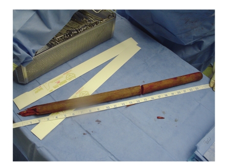
Put back together, the rod measured 63.5 centimetres.
Then the damage was repaired: the holes in the diaphragm and the stomach, the tears in the mesentery and the perforation of the rectum. Because the injuries were so extensive and the abdomen was full of wood splinters, the surgeons expected infection and chose a Hartmann’s procedure. The damaged lower bowel is closed off and the end of the colon is brought out through the abdominal wall as a colostomy, so that nothing passes the repaired rectum while it heals. They also closed the lining over the pelvis to seal it off from the rest of the abdomen, and left drains in the chest, the middle of the chest and the abdomen.
The whole operation took 3 hours and 52 minutes. It was carried out by a trauma surgeon, a cardiothoracic surgeon and three surgical residents.
Surviving the rod was the first part
In the surgical intensive care unit his pain was controlled with a pump he could press himself, and he was fed through a tube passed into his stomach. He developed a new irregular heart rhythm, atrial fibrillation, which was treated with amiodarone and beta blockers.
Two weeks after the operation he caught pneumonia in hospital, and it progressed to severe sepsis. He was treated with antibiotics and the full bundle of first-day sepsis measures, needed several new lines, a bronchoscopy and a tracheostomy, and the sepsis resolved.
There were other complications. His right arm was weak because of a complete brachial plexopathy, damage affecting the whole of the brachial plexus, the network of nerves that runs from the neck into the arm. He developed a hernia through the abdominal incision, and fluid collected in pockets around the right lung, which made it slow to wean him off the ventilator.
Two months after he arrived he was discharged, in good functional condition, to the hospital’s inpatient rehabilitation unit, and a week later he went home.
Closing the colostomy
Seventeen months after the injury his colostomy was reversed and the hernia repaired. A year after that he needed a second hernia repair. In all he went to the operating theatre three times over about two and a half years, and at the time of the report he was doing well.
Despite the wood splinters, the colostomy did its job: there were no infections inside the abdomen. The authors call the rod’s route fortunate, and say that handling it as little as possible was critical, both to keep any pressure it was putting on torn vessels and to limit blood loss.
An injury a novelist had already described
The authors’ review of the English-language literature suggests this is only the second reported case in a hundred years of an impalement through the pelvis, abdomen and chest being successfully treated. They describe a similar one from 1981. A 26-year-old woman jumped from a second-storey window of a burning building and landed on a tree. A branch entered through the anus, travelled up and to the right through the mesentery, tore the right lobe of the liver, split the joints between rib and cartilage of the eighth to tenth ribs on the right, and came out through the skin around the nipple. She, too, left hospital with a temporary colostomy, after eight weeks.
Impalement injuries have been recorded since ancient Egypt, and in the medieval period impalement became a common method of torture and execution. The authors point to the most famous description of it in literature, in the Nobel Prize winner Ivo Andrić’s novel The Bridge on the Drina, where an executioner drives a stake up through a man’s body so that it misses the vital organs and comes out near his right shoulder, deliberately, to keep him alive longer. That particular execution is fiction, though impalement itself was real. But the path Andrić described, up and to the right through the pelvis, abdomen and chest without fatal damage, is the one this patient survived, which suggests the novelist’s anatomy was sound.
There are no clear guidelines for operating on extensive impalement injuries, partly because every one is different. The authors’ recommendation is that objects like this should be removed in a major trauma centre, by surgeons who specialise in each region of the body the object has crossed, and with as little handling of the object as possible.
What this case teaches
A man who reportedly slipped in his bathtub arrived alert and stable, and the scans showed a wooden rod running from his rectum to his neck. Stable did not mean safe: an impaled object can be what is holding a torn vessel shut, so the team took him to theatre and opened both his chest and his abdomen before moving it, ready for the great vessels to bleed. They did not. The rod’s rounded end had apparently pushed past every major vessel on a 63.5-centimetre path through the bowel’s mesentery, stomach, diaphragm and the sac around the heart. It was cut in two inside the chest and each piece removed in the direction it had travelled, the injuries were repaired and the bowel protected with a colostomy. Pneumonia, sepsis, an irregular heart rhythm and weakness of his right arm followed, and he went home after two months and had the colostomy closed at 17 months. The principle is the same as for any impaled object: leave it in, and take it out only where everything it might be plugging can be reached.
Want the next case like this?
When a new case is written up, it comes to your inbox: the story, the diagram and what it teaches. No schedule and no filler. Unsubscribe in one click.
Free · no spam · your address is never shared · built with KitWas this case worth your time? Buy the next one a coffee ($3), or support MedicaseHub monthly.
Read the original paper: doi.org/10.1155/2009/361829
Adapted from Moncure M, Konie JA, Kretzer AB, DiPasco PJ and Braxton CC, “Survival Following Rectal Impalement through the Pelvic, Abdominal, and Thoracic Cavities: A Case Report,” Case Reports in Medicine 2009;2009:361829, from the University of Kansas Hospital, Kansas City. The presentation, imaging, operative findings and technique, the length and route of the object, the complications, the timeline and the outcome are as reported there, summarised in the author’s own words; the account of how the injury happened is as reported by the authors. The comparison with the 1981 case and with Ivo Andrić’s novel, and the recommendations on removal, are drawn from the paper’s discussion; the explanations of impalement, the anatomy, the Hartmann’s procedure and the treatments named are from standard references. The paper is published under a Creative Commons Attribution licence, and the two CT scans and the photograph of the rod are reproduced from it under that licence, unchanged. A photograph in the paper showing the patient’s neck and part of his face is not reproduced. The diagram is original to MedicaseHub, is simplified, and may be reused freely under CC BY 4.0. This article is not medical advice. If something is impaled in the body, do not pull it out: call emergency services, keep the object and the person as still as possible, and let surgeons remove it. Read the full disclaimer.
Related cases
- They cut the branch down. Nobody pulled it out.Another wooden object through the body, left in place until surgeons could reach what it had damaged.
- Wooden foreign body on CT: the scan said gasWhy wood can look like air on a CT scan, as the rod does here.
- The hand arrived still inside the machineAnother case where nothing was removed until the patient was in theatre.