They saved his ear by keeping it inside his belly
A road accident tore off a 22-year-old's left ear, and the wound it left was too badly damaged to sew it back on. So his surgeons stripped the skin from the ear, buried its cartilage under the skin of his abdomen to keep it alive, and three months later brought it back to his head.
- Specialty
- Otolaryngology, reconstructive surgery
- Patient
- Man, 22
- Mechanism
- Road traffic accident
- Injury
- Left ear completely torn off; the ear was found at the scene
- Problem
- Too much tissue lost around the ear, and no vessels to reconnect
- Day 1
- Ear cartilage stripped of skin and placed under the skin of the abdomen
- 3 months
- Cartilage taken out, alive and intact, and put back on his head
- 6 months
- Reconstruction complete
- Hearing
- Normal throughout
- day 0the accident
- day 1why it could not go back
- day 1into the abdomen
- 3 monthsout again
- 6 monthsthe finished ear
An ear, brought in separately
He was 22 when he was brought to the emergency department after a road traffic accident involving a motor vehicle. His left ear had been torn off completely. Behind where it had been there was a large wound, with a lot of skin and the tissue under it lost. The ear itself had been found at the scene and came to the hospital with him.
Apart from that he was well. There were no other injuries to his head, face or neck. The cartilage framework inside the severed ear was largely intact. His ear canal and eardrum looked normal, and a hearing test showed normal hearing in both ears: the injury had taken the outer ear, the part that collects sound, without reaching the parts that hear.
Why it could not simply be sewn back on
The best way to save a severed ear is to replant it under a microscope, joining its tiny arteries and veins to vessels at the edge of the wound so that blood flows through it again. When that is possible it gives the best result, because the patient gets his own ear back in its own shape.
Here it was not possible. Too much of the tissue around the ear had been torn away, and there were no suitable vessels left at the wound to join the ear's vessels to. An ear sewn on without a blood supply would simply die.
That left three options. Surgeons can carve a new ear framework from the patient's rib cartilage, a well-established technique with good long-term results, but it needs a second operation on the chest, considerable expertise and several stages over a long time. They can use a ready-made framework of plastic or silicone, which avoids the chest operation but is more likely to become infected or work its way out through the skin, especially in a wound that is already damaged. Or they can keep the patient's own ear cartilage alive somewhere else in his body until the wound has healed. The team chose the third.
Parking the cartilage under his skin
Within 24 hours of the accident he was in the operating theatre. The severed ear was cleaned, and the skin covering it was removed, leaving only the cartilage framework, the springy skeleton that gives an ear its shape. It was checked and found to be intact.
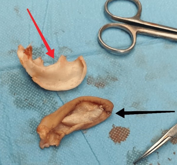
Then, under general anaesthetic, the surgeons made a small cut across the front of his abdomen and opened a pocket in the fatty layer just under the skin. They slid the cartilage into it the right way round, closed the wound over it in layers, and left a small suction drain to stop blood collecting. The drain came out on the third day.
Cartilage has no blood vessels of its own. It lives on nutrients that soak in from the tissue around it. Buried in the well-supplied fat of the abdominal wall, it could be kept alive by the tissue around it, and that is the whole idea of the technique, first described in 1972.
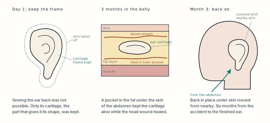
Want this diagram print-ready? Get it for $5, or all 42 in the diagram pack.
For the next three months he carried the frame of his left ear under the skin of his belly. The abdominal wound healed without infection, without the wound opening, and without the cartilage pushing through the skin. Meanwhile the wound behind his ear was dressed and allowed to heal on its own.
Taking it back out
Three months after the first operation, with both wounds healed and the patient in good health, the surgeons opened the abdominal pocket again under general anaesthetic and took the cartilage out.
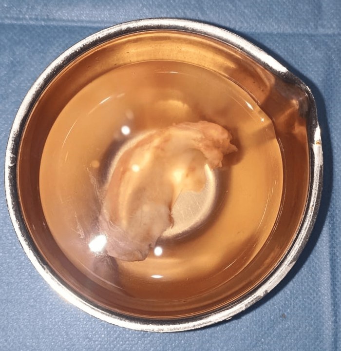
It had survived. The framework was intact and alive, with its curves still in place: the rim of the ear and the inner ridge just inside it. There was no sign that it had started to be absorbed, the main risk when cartilage is stored this way for too long, and no infection. What remained on his abdomen was a thin, well-healed scar.
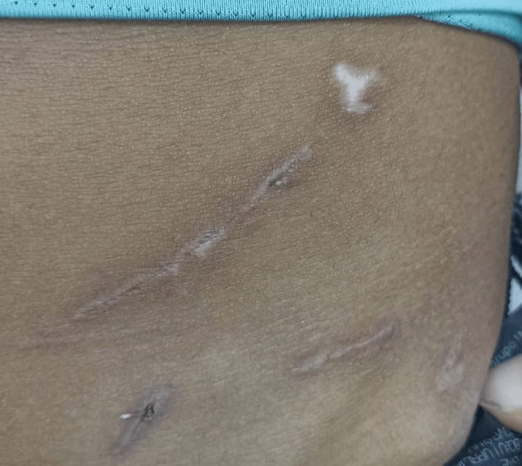
Back at the side of his head, the surgeons cut away the scar tissue that had formed where the ear had been and made a pocket with a good blood supply to receive the cartilage. They placed the framework in its proper position, held it with dissolving stitches, and covered it with skin moved in from the surrounding area and closed directly over it.
His own ear, slightly changed
Three months after the cartilage was put back, the reconstruction was complete, six months after the accident. There had been no wound problems, no collection of blood and no sign of the cartilage working its way out. The new ear sat naturally against the side of his face and stood out from the head as an ear should. Its shape was acceptable, with a mild distortion of the curve of its rim, which the authors note is a well-known and expected result of this technique. He was satisfied with how it looked and worked.
His hearing tests at three months after the final operation were unchanged from the day of the accident, and his eardrum was intact on both sides. The accident and the three operations had not affected his hearing.
Why the belly, and why three months
The abdominal wall is a good place to store cartilage because its fatty layer has a good blood supply to keep the cartilage alive, and the scar it leaves is easy to hide. Published series report cartilage kept this way for anywhere from six weeks to six months. Three months, the authors explain, is a compromise: long enough for the wound on the head to heal properly, but not so long that the stored cartilage starts to be absorbed and lose its shape.
The approach has its limits. The skin that once covered the ear cannot be saved, so the new covering comes from nearby, and the fine curves of the ear are usually a little blurred, as they were here. But it gives the patient back his own cartilage in its own shape, without surgery on the chest and without an artificial implant, and it needs no microsurgery. That, the authors point out, makes it particularly useful where microsurgical teams and equipment are not available.
For anyone who finds a severed ear, finger or other body part after an accident, the first steps are the same: call for emergency help, and bring the part to hospital with the patient. Here, finding the ear at the scene is what made everything else possible.
What this case teaches
A 22-year-old lost his left ear in a road accident, and the wound behind it was too damaged, with no vessels to connect to, for the ear to be sewn back on. Within a day his surgeons removed the skin from the severed ear and buried its cartilage framework under the skin of his abdomen, where the surrounding tissue kept it alive. Three months later, with the head wound healed, they took the cartilage out intact and placed it back on the side of his head under skin moved from nearby. Six months after the accident he had his own ear again, with a slightly blurred rim, and his hearing was normal throughout. When a severed part cannot be reconnected straight away, the body itself can sometimes keep it alive until it can.
Was this case worth your time? Buy the next one a coffee ($3), or support MedicaseHub monthly.
Next case · MC-038 They operated on the hand before they operated on himA machine took his hand off through the forearm. One team worked on the hand on a side table while the other prepared him, and blood was crossing it again four hours later.Read it →
Want the next case like this?
When a new case is written up, it comes to your inbox: the story, the diagram and what it teaches. No schedule and no filler. Unsubscribe in one click.
Free · no spam · your address is never shared · built with KitRead the original paper: doi.org/10.7759/cureus.112141
Adapted from Taleuan A, Lakhal A, Squalli Houssaini ME and Oudidi A, “Auricular Cartilage Preservation Through Abdominal Subcutaneous Implantation Following Traumatic Ear Amputation: A Case Report,” Cureus 2026;18(7):e112141, from Al Amir Sultan Hospital, Errachidia, and the Centre Hospitalier Universitaire Hassan II, Fez, Morocco. The injury, the examination and hearing tests, the reasons replantation was not possible, the operative technique, the timeline and the outcome are as reported there, summarised in the author’s own words; the comparison with rib cartilage and artificial frameworks, the history of the technique and the reasoning behind the three-month interval are drawn from the paper’s introduction and discussion, and the explanation of how cartilage is nourished is from standard references. The paper is published under the Creative Commons Attribution licence (CC BY 4.0). The three photographs are reproduced from its Figures 3, 8 and 9 under that licence, cropped; the authors state that consent for open-access publication was obtained or waived. The photographs of the original wound and of the operations, which show open wounds, and the final photograph, which shows the patient’s face, are not reproduced. The diagram is original to MedicaseHub, is simplified, and may be reused freely under CC BY 4.0. This article is not medical advice. Read the full disclaimer.
Related cases
- They operated on the hand before they operated on himA severed part that could be reconnected, and the race to do it in time.
- Half his face was torn away. It was all put back in one operation.Another injury where saving the patient's own tissue made all the difference.
- Replanting an arm: the clock, and the harder questionHow long a severed part can survive, and why it depends on what it is made of.