The silicone in her arm came from her breast implant
She had silicone implants placed in both breasts in 1987. After a car crash, her left breast slowly swelled, and nearly three years later an MRI raised concern for cancer. Surgery found a ruptured implant there and about 600 millilitres of organised haematoma, old blood that had partly turned into firm tissue. On the right, silicone from the other implant had escaped into her armpit and upper arm.
- Specialty
- Plastic surgery
- Patient
- Woman, 65, with high blood pressure
- Implants
- Silicone, both breasts, placed in 1987
- Onset
- Swelling of the left breast that began after a car crash
- Delay
- Nearly three years from the crash until she was seen
- Left breast
- Large, firm, fixed mass low in the breast, red skin over it
- Right arm
- Fluctuant, non-tender lump, 8 × 4 cm, inner upper arm
- MRI
- Masses behind both chest muscles, concerning for cancer; silicone extending into the right armpit and arm
- Biopsy
- Organised haematoma, benign
- Found
- Both implants ruptured; organised haematoma of about 600 mL on the left and 200 mL on the right
- Follow-up
- Four units of blood after surgery; well at one month
- 1987the implants
- crashthe swelling begins
- 3 yearsa mass in each place
- scanit looked like cancer
- theatreboth implants had failed
- 1 monththe cost of waiting
What can happen to an implant over the years
She was 65 when she was seen, with high blood pressure, and she had had silicone implants placed in both breasts in 1987.
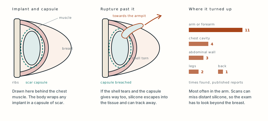
Every implant ends up inside a capsule. The body treats it as a foreign object and walls it off with a thin layer of scar tissue, which is normal and usually goes unnoticed. Sometimes that capsule tightens around the implant. This is called capsular contracture, and when she was examined she had marked contracture on both sides.
Implant shells can also tear. When the rupture stays inside the capsule, patients often have no symptoms, although some notice tightness, a change in shape or pain. When silicone gets past the capsule, it comes into contact with the surrounding tissue, which can react to it with a chronic inflammation called a granuloma. Rarely, the silicone travels much further than that and turns up as a lump under the skin somewhere else in the body.
A breast that changed after a car crash
Her symptoms began after a car crash, three years before she was seen at the hospital that wrote up her case.
Over that time the lower part of her left breast slowly swelled, and the skin over it became painful and tender. She first went to another hospital, but for insurance reasons she could not get definitive treatment, and nearly three years had passed since the crash by the time she arrived.
Throughout that time she stayed stable, with no symptoms of anaemia, and she said she was not taking any medicine that thins the blood or affects platelets. That last detail matters because of what was later found in her breast.
One breast, and one arm
Both breasts showed marked capsular contracture. Low in the left breast was a large, firm mass that did not move, and the skin over it was red, with increased blood vessels. On the right there was extensive fullness across the upper part of the breast, extending into the armpit.
Then there was her right upper arm. On its inner side, over the brachialis, one of the muscles that bend the elbow, was a lump measuring 8 by 4 centimetres that felt fluid-filled when pressed. It was not tender, and the skin over it looked normal.
She reported none of the general symptoms, such as fevers, night sweats or weight loss, that can come with cancer, and no broken skin, nipple discharge or pulling in of the nipple. Her last mammogram, about three years before, had shown no sign of cancer, and she had never had breast cancer. All of that was reassuring, but it did not rule cancer out.
The scan raised concern for cancer
MRI of the breasts showed large, uneven, oval masses behind the chest muscles on both sides, and they were reported as concerning for cancer. There was also a collection of fluid in the left breast.
The images of the right side of her chest showed something else too: silicone outside its capsule, extending from the right breast into the armpit and the arm. That put escaped silicone in the same arm as the lump.
Because of the concern for cancer, a core biopsy was taken from the mass low in the left breast. It was benign. It showed an organised haematoma: a collection of blood old enough to have partly turned into firm tissue.
Bleeding around a breast implant is a known complication, but it is usually seen within the first three days after the operation. A haematoma appearing more than six months later is rare enough that one review found only 31 such patients reported after cosmetic breast augmentation. Hers was one of those rare late cases.
Two ruptures, and silicone in the arm
She was taken to theatre to have both implants removed together with the whole of each capsule, an operation called a total capsulectomy.
The right implant had ruptured completely, and silicone had been pushed out through the right armpit and into the upper arm. An organised haematoma of about 200 millilitres, containing fibrin and silicone granulomas, was cleared from the right breast. The silicone in the arm was taken out by hand through a cut in the upper arm, along the groove beside the biceps.
The left implant, a 300 cc implant, had ruptured too, and lay inside a fibrous capsule. The larger problem was on this side: an organised haematoma of about 600 millilitres, removed in pieces from the lower part of the breast. The red, congested skin over it was removed as well.
Specimens from both sides went to the pathologists, and both showed benign haematoma with scattered granulomatous reaction. Specific staining for anaplastic large cell lymphoma, a rare lymphoma linked to breast implants that can appear as a late swelling around one, was negative.
The wounds were closed directly, with a drain left in each breast pocket.
The cost of waiting
Removing the haematoma caused significant blood loss, and afterwards she was given four units of packed red blood cells. Apart from that she recovered well, and when she was seen one month later she had no further complaints or complications.
The authors are direct about the delay. In their view, the three years between the crash and the operation likely allowed the haematoma to expand, and they consider that this is likely why she needed a far more invasive operation and a blood transfusion.
They also suggest a possible mechanism. The silicone that escaped at the start may have set up a chronic granulomatous reaction, which then led to repeated fresh bleeding on top of old, what they call an acute-on-chronic haematoma. They put this forward as a possibility, not as something the case proves.
Their other point is about patients. People with implants should know the signs of a rupture and come back to be seen if they notice them.
Where escaped silicone goes
The authors searched the medical literature for other cases of silicone travelling away from a ruptured implant and found 19 case reports describing 20 patients. Across those reports and their own case, the median age at presentation was 48, ranging from 21 to 76, and the median time since the implants were placed was 10 years, ranging from 1 to 30.
The arm or forearm was the most common site, recorded 11 times. The chest cavity followed with 4, then the abdominal wall with 3, the legs with 2 and the back with 1. Of the patients, 67% had a documented injury to the chest before they presented.
Two of the older reports the authors cite describe silicone escaping after a closed capsulotomy, a technique once used to break a contracted capsule by squeezing the breast from outside. Another describes silicone that had infiltrated a nerve and caused a constrictive neuropathy.
Many of the implants in the series dated from the 1970s to the early 1990s, before highly cohesive implants were developed. Whether that change affects how often silicone travels is, as the authors note, still an open question.
Their practical point is about examination. Imaging can miss silicone that has travelled far from the breast, so when a rupture is suspected, the physical examination needs to be thorough and go beyond the breast itself.
What this case teaches
Two problems were found in the same woman. On the left, a breast that began to swell after a car crash went untreated for nearly three years, and by then it held a ruptured implant and about 600 millilitres of organised haematoma, with red skin over it and an MRI that raised concern for cancer. On the right, silicone from a completely ruptured implant had been pushed through the armpit into the upper arm, the same arm where a fluctuant lump could be felt. A biopsy settled the question of cancer, the MRI showed the silicone beyond the capsule, and one operation removed both implants, both capsules, the haematomas and the silicone in the arm. In the authors’ view, the delay likely cost her a far more invasive operation and a blood transfusion. The lessons are just as plain: a breast with an implant that changes after an injury to the chest needs investigating, a lump in the arm of someone with implants may have come from them, and the examination has to go further than the breast.
Read the original paper: doi.org/10.1097/GOX.0000000000001849
Adapted from Dean RA, Glener AD, Thomas AB, Glener SR, Kurtovic S and Erdmann D, “Silicone Migration and Late Hematoma following Silicone Implant Rupture: Case Report and Literature Review,” Plastic and Reconstructive Surgery Global Open 2018;6(8):e1849, from the Division of Plastic, Maxillofacial, and Oral Surgery, Department of Surgery, Duke University Health System, Durham, North Carolina. The history, examination, imaging, biopsy, operative findings, volumes, pathology, transfusion and follow-up are as reported there, summarised in the author’s own words rather than reproduced. The literature review figures, the authors’ view of the delay, the suggested mechanism and their points on examination and patient education are drawn from that paper; the mentions of closed capsulotomy and nerve involvement come from the titles of reports it cites, and the background on capsules, capsular contracture, granulomas, organised haematoma and implant-associated lymphoma from standard references. The paper is published under a Creative Commons Attribution Non-Commercial No-Derivatives licence, so its clinical photographs, MRI images and video are not reproduced here. The diagram is original to MedicaseHub, is simplified, and may be reused freely under CC BY 4.0. This article is not medical advice. A change in the shape, size or feel of a breast with an implant, especially after an injury to the chest, or a new lump in the armpit or arm, should be checked by a doctor. Read the full disclaimer.
Find these cases useful? Support MedicaseHub on Patreon.
One case, written up properly, when there is one worth sending
No schedule and no filler. When a case turns out to have something in it that is worth the time to explain, it goes out. Unsubscribe in one click.
Free · no spam · your address is never shared · built with KitRelated cases
- It was never cancer. It still had to come out.Another mass that was not cancer and still needed an operation.
- Wooden foreign body on CT: the scan said gasA scan that misread what was in the tissue, and the clinical sign that corrected it.
- The steroid was for his muscles. It killed the skin above them.Something put into the body for how it looks, and the damage it did to the tissue around it.