The lion was a gift. Three weeks later it attacked him.
A 17-year-old was attacked by the lion his father had given him as a pet. He arrived at hospital two hours later in shock, with his scalp torn loose almost all the way round his head and a broken skull with air inside it. Five days later he went home.
- Specialty
- Neurosurgery, plastic surgery, trauma
- Patient
- Male, 17, with asthma
- Mechanism
- Attack by a 5-year-old lion kept as a pet
- To hospital
- 2 h, brought by his father
- Head
- Scalp torn loose nearly all round; skull broken and pushed in above the left ear, with air inside
- Also
- Deep wound beside the left eye, torn right ear, deep bites to the chest and both arms, shock from blood loss
- Operation
- Skull and its lining repaired, scalp laid back and closed in layers
- Hospital
- Home on day 5
- Two weeks later
- Wounds healed, scalp alive, brain function intact
- 0 hthe attack
- 2 harrival
- day 0the operation
- day 5home
- +2 weekshealed
A pet that was not yet home
The lion was five years old. According to the boy’s father, he had given it to his son as a gift three weeks earlier, to be kept as an exotic pet.
It was not living with the family yet. Until the boy had finished preparing his own facilities for it, the lion was being housed at a friend’s residence, because the friend already kept other exotic animals and had enclosures suitable for them. The attack happened during one of the boy’s visits there.
The report does not describe the attack itself, only what it left behind. Two hours later his father brought him to the emergency department of Hamad Medical Corporation in Doha, Qatar.
Awake, and in shock
He was fully conscious, with a Glasgow Coma Score of 15 out of 15, the highest possible. But his blood pressure was low, 98/60, and his heart was racing at 120 beats a minute: the signs of shock from blood loss.
The first examination found four main injuries. His scalp had been torn loose from the skull almost all the way round his head, and it was bleeding heavily. There was a large wound at the outer side of his left eye, in which muscles had been cut through. There were deep bite wounds over his chest and both arms. And his right ear had been torn through its full thickness, exposing the cartilage inside.
The scalp is built in five layers: the skin, a dense fatty layer carrying its blood vessels, a tough sheet of tendon called the galea, a thin layer of loose tissue, and the pericranium, the membrane on the surface of the bone. The loose layer is a natural plane of weakness. When the scalp is pulled hard enough, it peels away there, all its upper layers together, leaving the bone beneath covered only by its thin membrane. Surgeons call this a degloving injury, because the scalp comes off the way a glove comes off a hand.
A CT scan of his head showed something more serious underneath. The temporal bone, the part of the skull above and around the left ear, was broken into several pieces, and they had been pushed inwards. The fracture was open to the wound, and there was air inside his skull, a finding called pneumocephalus. Air does not get inside the skull unless something has broken through it.
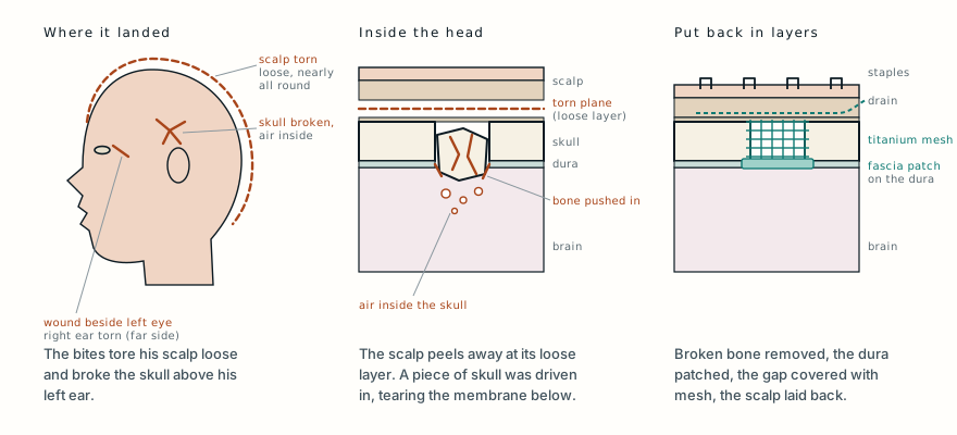
He was stabilised quickly and taken straight to the operating theatre, where two teams would work on him in turn: neurosurgeons first, then plastic surgeons.
From the inside out
Large parts of the scalp had been torn away through its full thickness, and some areas of it had a compromised blood supply. Underneath, the neurosurgeons removed the pieces of broken bone that were no longer viable. Below the fracture they found a tear in the dura, the tough membrane that wraps the brain and keeps the fluid around it sealed in.
They repaired the dura with a patch of temporalis fascia, the strong sheet of tissue that covers the chewing muscle at the temple, taken from the boy himself. Then they rebuilt the gap in his skull with titanium mesh.
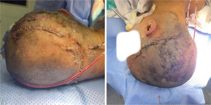
The plastic surgeons took over. They debrided and irrigated every bite wound, cutting away damaged tissue and washing the wounds out. Then they laid the scalp back in its place and closed it in layers, without tension, so as not to squeeze off its blood supply: dissolving stitches for the deeper layers, finer dissolving stitches for the more delicate areas, and staples for the skin. Two drains were left under the scalp, so that blood could not collect beneath it. No blood vessels were rejoined under a microscope; the scalp was put back and stitched, and it survived on the blood supply it still had.
The cut muscles beside his left eye were stitched back together. The ear was repaired by realigning its cartilage. The deep wounds on his chest and arms were washed out, cleaned and closed.
PhotographThe back and side of his head right after the operation, with the scalp stapled back in place. Tap to show.
Antibiotics, tetanus, and rabies
He spent two days in the trauma intensive care unit and was then moved to a step-down unit.
A lion’s mouth carries many kinds of bacteria, and its teeth push them deep into the tissues. He was given a broad-spectrum intravenous antibiotic, piperacillin-tazobactam, and protection against tetanus. After a discussion with the infectious disease specialists, he was also started on rabies post-exposure prophylaxis.
His wounds were inspected every day. Physiotherapy started early to keep his arms moving. On the fifth day after the operation the drains were removed, and he went home.
The scalp held
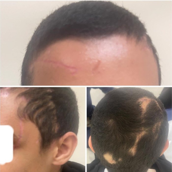
At his review two weeks later, all the surgical sites had healed well. The scalp that had been laid back was alive and well perfused, his hairline was preserved, and his neurological function was intact. The text of the report describes no areas of hair loss; the caption of the photographs mentions small scattered patches without hair, which can be seen along some of the scars.
The report does not describe how his left eye, or his vision, recovered.
Why big cats do this much damage
Attacks by large predators are rare, but they are often catastrophic. The authors cite a global review of 5,089 incidents involving large predators in which 32% were fatal, and most of the survivors were left with significant injuries. Lions alone are estimated to kill about 250 people a year worldwide, and many more non-fatal attacks go unreported.
A lion’s bite and claws can drive through soft tissue, break bone and cause heavy bleeding. Injuries to the head, including skull fractures, are common in these attacks, and the authors cite a study by Mowatt and colleagues that found head and neck injuries among the most likely to be fatal.
Infection is the second danger. Bites from big cats carry a mixture of bacteria from the animal’s mouth, including organisms such as Pasteurella multocida and Capnocytophaga canimorsus, and the bites go deep. That is why every such wound is cleaned and washed out, why antibiotics are started early, and why tetanus protection is given. Rabies has been documented in lions, and the authors note that World Health Organization guidelines call for wound care, rabies vaccine and rabies immunoglobulin after exposure.
The authors end with a point that has nothing to do with surgery. In their view, the trend of keeping lions, tigers and other large carnivores as private pets is a public health problem, and incidents like this one are largely preventable. They call for stronger regulation of exotic animal ownership, public education, veterinary oversight and emergency preparedness.
What this case teaches
A 17-year-old was attacked by a five-year-old lion that his father had given him as a pet three weeks earlier. He arrived awake but in shock, with his scalp torn loose almost all the way round, a broken skull pushed in above his left ear with air inside it, a deep wound beside his left eye, a torn ear and deep bites to his chest and arms. Neurosurgeons removed the broken bone, patched the torn lining of the brain with tissue from his own temple and covered the gap with titanium mesh; plastic surgeons then laid his scalp back and closed it in layers. Antibiotics, tetanus protection and rabies prophylaxis followed, and he went home on the fifth day. Two weeks later his scalp had survived and his brain function was intact. His surgeons credit fast, coordinated care for that outcome, and add that attacks like this one are largely preventable.
Was this case worth your time? Buy the next one a coffee ($3), or support MedicaseHub monthly.
Next case · MC-046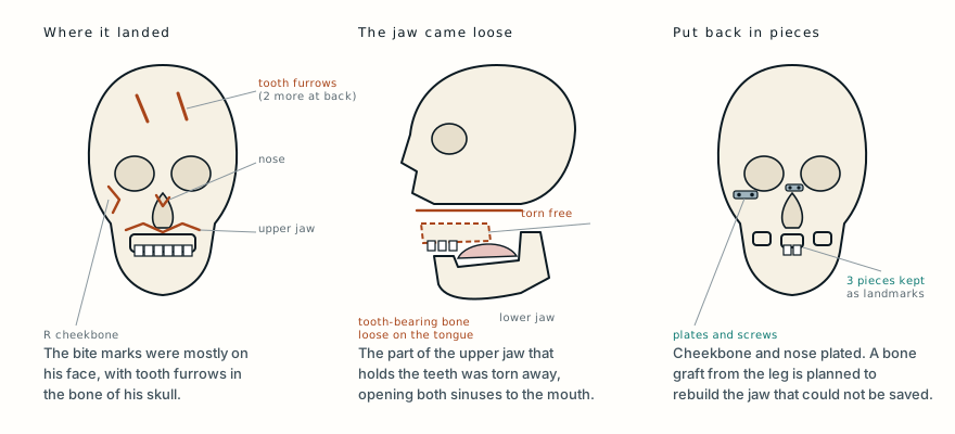 The bear went for his face, and left its tooth marks in his skullA man gathering pine nuts alone in a Siberian forest was attacked by a bear. He walked away and reached hospital nine and a half hours later, with his upper jaw torn loose and tooth furrows in his skull.Read it →
Want the next case like this?
When a new case is written up, it comes to your inbox: the story, the diagram and what it teaches. No schedule and no filler. Unsubscribe in one click.
Free · no spam · your address is never shared · built with KitRead the original paper: doi.org/10.5339/qmj.2025.123
Adapted from Omari RY, Al Alawi K, Abdelrahman R, Alyazji Z, Elsharkawy M and Hammouda A, “Multidisciplinary reconstruction of complex cranio-scalp trauma in a patient with a domestic lion attack,” Qatar Medical Journal 2025;2025(4):123, from Hamad Medical Corporation, Doha. The background to the attack, which is the father’s account as reported by the authors, the examination and scan findings, the operations, the hospital course and the follow-up are as reported there, summarised in the author’s own words; the figures on predator attacks and the points on infection, rabies and exotic pets are those the authors cite in their discussion, and the explanations of the anatomy are from standard references. The paper is published under the Creative Commons Attribution licence (CC BY 4.0), and the authors report written consent from the patient’s parents to publish the case and images. The two photographs here are reproduced from Figures 2 and 3 under that licence, one of them cropped. The photographs of his wounds on arrival are not reproduced. The diagram is original to MedicaseHub, is simplified, and may be reused freely under CC BY 4.0. This article is not medical advice. Read the full disclaimer.
Related cases
- The bear went for his face, and left its tooth marks in his skullA bear this time, and tooth furrows in the bone of the skull.
- Half his face was torn away. It was all put back in one operation.Another torn flap of the head that survived because it was put back fast.
- The bone plate was screwed on outside his legA crocodile bite, and why animal wounds are treated as contaminated.