The bear went for his face, and left its tooth marks in his skull
A 42-year-old man was gathering pine nuts alone in a Siberian forest when a bear attacked him. It bit through the arm he raised to protect himself, pinned him down and went for his face. He stayed conscious, walked back towards his camp, and reached hospital nine and a half hours later with his upper jaw torn loose.
- Specialty
- Maxillofacial and plastic surgery, trauma
- Patient
- Man, 42
- Mechanism
- Attack by a wild bear while gathering pine nuts
- To hospital
- 9 h 30 min, by helicopter
- Face
- Broken cheekbone, nose and upper jaw; tooth-bearing bone torn loose
- Also
- Open fractures of the left arm and hand, bites to the trunk and limbs, shock from blood loss
- Operation
- Wounds cleaned, bones plated, an eye muscle reattached
- Hospital
- Discharged on day 23
- Now
- Cannot chew; a bone graft from the leg is planned
- 0 hthe attack
- 9 h 30 minarrival
- day 1the operation
- day 23home
- 3 monthswhat was lost
Alone in the forest
One morning in September 2023, in a forest in southern Siberia, a 42-year-old man was harvesting pine nuts on his own when a wild bear attacked him.
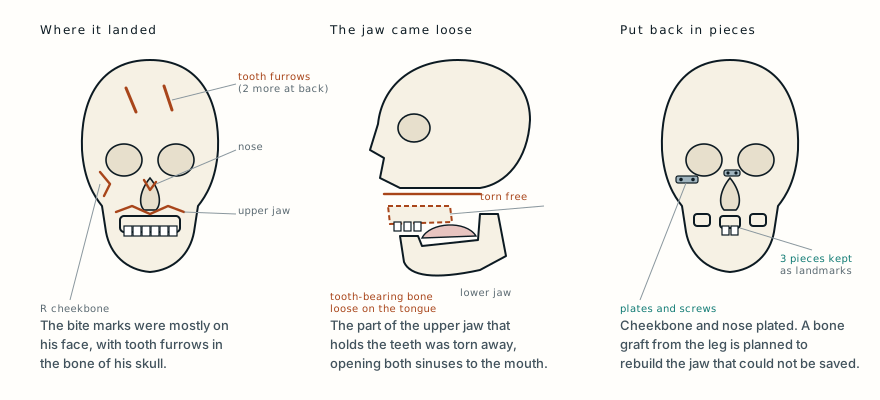
According to his account, he raised his left arm to cover his face and the bear bit through it. Then it pinned his body to the ground with a paw and began biting his face. The authors write that it then tried to open his skull: there were four furrows from its canine teeth in the bone, two on the forehead and two at the back of the head. He stayed conscious throughout. When he stopped resisting, the bear left.
He waited about ten minutes, got up on his own, checked that the animal had gone, and set off towards his camp. A helicopter rescue service was called. Nine and a half hours after the attack he arrived at the Republican Clinical Hospital in Abakan.
A face full of forest
A CT scan of his whole body was done straight away, and five specialists examined him. The intensive care doctor found him in shock from blood loss. The trauma surgeon found open fractures of his left arm: the lower end of the upper arm bone was shattered, the ulna in the forearm was broken, and so was a bone at the base of the thumb. The neurosurgeon found a head injury, though the scan showed no damage to the brain itself. The general surgeon found bite wounds over his trunk and limbs, but no injury to the organs inside. His face was the worst of it, and he was admitted to the maxillofacial and plastic surgery department.
The wounds on his face were full of sand, hay, pine needles, grass, leaves and dead insects. On the right cheek a flap of tissue had been torn open into his mouth. In the bottom of it the surgeons could see a partly severed facial nerve, the parotid salivary gland, torn facial muscles, and fragments of his upper jaw with teeth still in them. His right cheekbone and his nose were broken into displaced pieces. On the left there was a crescent-shaped wound over the other parotid gland.
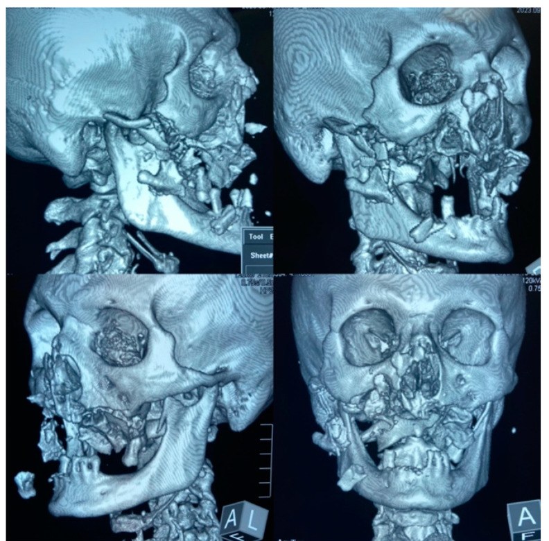
Inside his mouth the damage was even more striking. The lining of the palate was torn in several places. The tooth-bearing part of his upper jaw, the alveolar process, had been torn away from the rest of the skull and was lying loose on his tongue. With it gone, both maxillary sinuses, the air spaces in the cheekbones, now opened straight into his mouth. The front section, still holding his two upper front teeth, had been twisted round and pushed into his left cheek.
The CT also showed a broken arch of the right cheekbone, multiple fractures of the walls of the right maxillary sinus, a fracture of the floor of the left one, a broken projection of the lower jaw on the left, and a great deal of air trapped under the skin of his face and scalp. The surgeons classified the fractures of the upper jaw as Le Fort II on the right and Le Fort I on the left, the standard patterns in which the midface breaks away from the skull. One of the muscles that moves his left eye, the superior rectus, had been torn loose, the duct that drains the right parotid gland was torn, and the right side of his face was partly paralysed.
He was given a rabies vaccine and tetanus protection.
Cleaning, then rebuilding, layer by layer
The arm came first. Its wounds were cleaned, the bones were held in line with temporary wires and the arm was put in plaster.
Then the face. Before anything could be repaired it had to be cleaned, and the surgeons removed leaves, pine needles, grass, broken teeth from both jaws and small fragments of bone from the wounds.
He was started on two intravenous antibiotics for a week. Because the broken right cheekbone could be seen clearly through the existing wound, the surgeons put its pieces back in place and fixed them with small titanium plates and screws. The broken bones of the bridge of the nose were set and plated in the same way.
The loose pieces of the upper jaw were taken out of the soft tissues of his face. Three of them were kept because they were large and would serve as anatomical landmarks for the reconstructions still to come: the two rounded back ends of the upper jaw, and the front segment with his two front teeth. These were fixed back in place with plates and screws. Crushed and dying lining of the mouth was cut away, the sinuses were packed with antiseptic gauze led out through the nose, and the lining of the cheeks was brought over the fixed fragments. Sewing the lips closed then separated the inside of the mouth from the wounds of the face.
One small wound, 0.7 centimetres long, near his left upper eyelid, hid something unexpected. Hanging out of it was what looked like a tendon attached to a muscle. When the surgeons pulled gently on its end, his eye moved. It was the torn end of the superior rectus, one of the muscles that turn the eye. They stitched it to the firm plate inside the upper eyelid. The next day both his eyes moved normally and together in every direction.
Two-thirds of the operating time went on closing the wounds of his face in layers: the capsules of the salivary glands, the muscles and the fat under the skin with dissolving stitches, and then the skin. During the operation he was given 686 millilitres of red blood cells and 580 millilitres of plasma, and another 1,350 millilitres of red cells afterwards in intensive care.
Three weeks in hospital
He went to intensive care on a ventilator and was breathing on his own by the next morning. He was fed through a tube into his stomach to keep food away from the wounds in his mouth, and given atropine before meals to reduce saliva, because a torn salivary gland can leak saliva out through the skin of the face as a fistula.
He moved to the ward on day 8. Salivary fistulas did form over the wounded parotid glands, and they closed on their own by day 15. Some stitches in the mouth gave way over the upper jaw, and on day 16 the lining of the mouth was repaired again to stop holes forming between the mouth and the sinuses. On day 23 he went home.
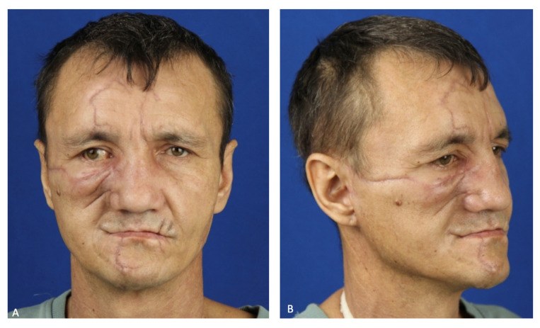
PhotographHis face two and a half months after the attack. Tap to show.
The bone that did not survive
From late November his scars were treated with a laser and with injections of a collagen gel to help them settle.
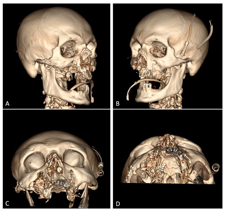
In early December he was readmitted. Parts of his upper jaw had become chronically infected and the bone was dying, a condition called osteomyelitis: the bone of the tooth-bearing ridge lay exposed in his mouth, yellow-grey, under red, inflamed lining. On 13 December the surgeons removed the dead bone with an ultrasonic instrument and took out the plates and screws on the right back end of the jaw.
At the time of the report he had completely lost the ability to chew, and the right side of his face moved poorly. The next stages were planned: restoring his ability to chew, rehabilitating the injured facial nerve, and transplanting bone from his fibula, the thin bone of the lower leg, to replace the missing part of the upper jaw so that dental implants could later be fitted. His left elbow had stiffened after the fractures, and he had a further operation on it, including removal of the head of the radius.
Why bears go for the face
Bear attacks are rare. The authors cite a United States National Park Service figure of one attack for every 2.7 million visits to protected areas. They also cite studies showing that about half of bear attacks happen during leisure activities in forests, such as picking berries or mushrooms, that almost all victims are adults, most of them men, and that most are alone, as this man was. Most attacks happen by day, and one study from Iran found that most were defensive, a reaction to a surprise encounter.
When a bear does attack, the head and face are the most common target. In the studies the authors review, most victims had facial injuries, the cheekbone and eye socket were among the bones most often broken, and most survivors were left with scars or a changed face that needed more surgery later. This case followed that pattern closely: the face took the worst of the attack, the bone of the upper jaw could not all be saved, and the reconstruction was still continuing months later.
Wounds from animal bites are contaminated, and these were full of soil and plant material. That is why every bear-attack victim needs thorough cleaning of the wounds, antibiotics, and protection against tetanus and rabies, and why infection of damaged bone can still appear weeks later, as it did here.
What this case teaches
A man gathering pine nuts alone in a Siberian forest was attacked by a bear that bit through his arm and then his face, leaving tooth furrows in the bone of his skull. He walked away, and reached hospital nine and a half hours later in shock, with his cheekbone, nose and upper jaw broken and the tooth-bearing part of the jaw lying loose on his tongue. His surgeons cleaned out pine needles, leaves, grass and his own broken teeth, plated what could be saved, reattached a torn eye muscle they found hanging from a small eyelid wound, and closed his face layer by layer. He went home after 23 days. But part of his upper jaw later died from infection, and at the time of the report he could not chew, with a bone graft from his leg still to come. Surviving a bear attack is often only the beginning of a long reconstruction, and the face, the bear's usual target, bears most of it.
Was this case worth your time? Buy the next one a coffee ($3), or support MedicaseHub monthly.
Next case · MC-040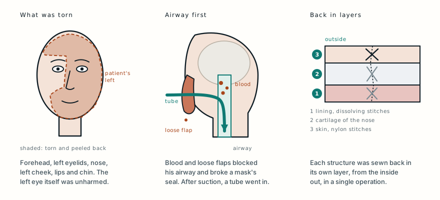 Half his face was torn away. It was all put back in one operation.A motorcyclist arrived with the left side of his face torn and peeled back, and his own tissue blocking his airway. Surgeons put every layer back in a single operation, and two weeks later he went home.Read it →
Want the next case like this?
When a new case is written up, it comes to your inbox: the story, the diagram and what it teaches. No schedule and no filler. Unsubscribe in one click.
Free · no spam · your address is never shared · built with KitRead the original paper: doi.org/10.3390/healthcare12050542
Adapted from Mellin R, Velichko E, Maltseva L, Dydykin S and Vasil’ev Y, “Polytrauma Caused by a Bear Attacking a Human with a Benign Outcome,” Healthcare 2024;12(5):542, from the G. Ya. Remishevskaya Republican Clinical Hospital, Abakan, and Sechenov University, Moscow. The account of the attack, which is the patient’s as reported by the authors, the examination and scan findings, the injuries, the operations, the hospital course and the later complications are as reported there, summarised in the author’s own words; the statistics on bear attacks are those the authors cite in their discussion, and the explanations of the anatomy and treatments are from standard references. The paper is published under the Creative Commons Attribution licence (CC BY 4.0), and the authors report written consent from the patient to publish his history and photographs. The two CT images and the photograph here are reproduced from Figures 1, 4 and 11 under that licence, one of them cropped. The photographs of his open wounds, of the material removed from them and of the operations are not reproduced. The diagram is original to MedicaseHub, is simplified, and may be reused freely under CC BY 4.0. This article is not medical advice. Read the full disclaimer.
Related cases
- Half his face was torn away. It was all put back in one operation.Another face torn open, and why so much of it could be put back in one sitting.
- He could breathe sitting up, but not lying downAnother midface broken away from the skull in the classic Le Fort pattern.
- The bone plate was screwed on outside his legA crocodile this time, and a contaminated open fracture of the leg.