Thirty-five operations could not stop the tumour. A new face did.
A young Polish woman with neurofibromatosis had a tumour growing through her whole face. After more than 35 operations in 24 years it was still threatening her speech, her eating and her sight. At 28, after seven years on the waiting list, she received the face, ears, eyelids and scalp of a 19-year-old donor.
- Specialty
- Reconstructive surgery, transplantation
- Patient
- Woman, 28
- Condition
- Neurofibromatosis type 1, with a plexiform neurofibroma through the whole face
- Before
- More than 35 operations in 24 years; left side of the face paralysed
- Waiting list
- June 2006 to December 2013
- Donor
- Woman, 19, brain dead after a car accident
- Transplanted
- Face, both ears, both eyelids and the scalp: over 75% of the soft tissue of the head
- Operation
- 24 hours, 27 units of blood
- At 1 year
- Near-complete smile, mouth closure 75%, feeling over 90%
- 24 yearsthe tumour
- Dec 2013the transplant
- weekswaking up to it
- 1 yeara smile
A tumour that would not stop
Neurofibromatosis type 1 is a genetic condition that affects about one person in every 2,500 to 3,300. It is inherited in a dominant pattern, so one altered copy of the gene is enough to cause it. One of its features is the neurofibroma, a benign tumour that grows from the sheaths of nerves. Most are small lumps in the skin. The plexiform type is different: it grows along the branches of a nerve, infiltrating everything around it, and it can become enormous.
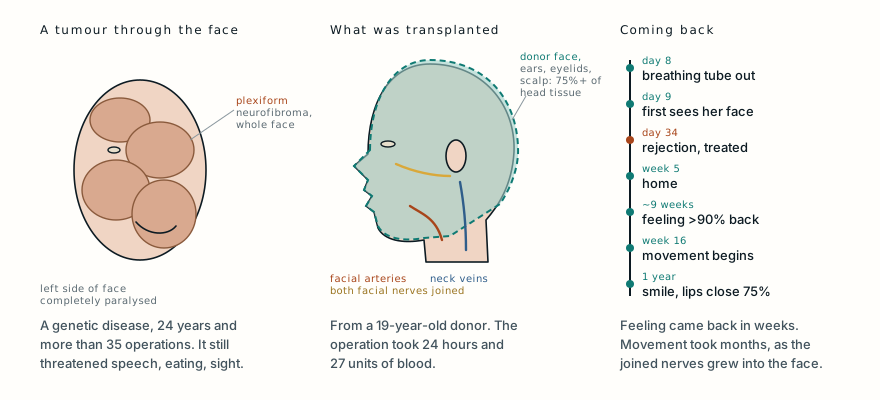
This woman’s disease was limited to her head, and a plexiform neurofibroma had spread diffusely through the upper, middle and lower parts of her face, distorting all of it. Over 24 years she had more than 35 operations to remove or reduce it. In the last three years before her transplant, her condition got markedly worse, with serious problems speaking, eating and seeing. The left side of her face was completely paralysed and the right side partly, probably because the tumour had infiltrated the ends of her facial nerves.
Conventional reconstruction moves a patient’s own tissue to rebuild what is missing. For a tumour that had invaded the whole face, there was nothing healthy left to rebuild from. Her surgeons at the Maria Skłodowska-Curie Institute cancer centre in Gliwice, Poland, considered her for a face transplant, and in June 2006 she was placed on the national transplant waiting list. She waited more than seven years.
Two operating rooms, 24 hours
On 4 December 2013 a donor was found: a 19-year-old woman who had been declared brain dead after a car accident, with her heart still beating. She was matched to the recipient on several clinical and biochemical characteristics, and tests showed no antibodies in the recipient against the donor’s cells. The face was the first tissue taken from the donor, before her other organs.
The operation ran in two connected operating rooms. In one, the surgeons removed the donor’s face. In the other, they removed the tumour from the recipient: all the soft tissue of the upper, middle and lower face. They found both facial nerves where they leave the skull, below the ear, and prepared the arteries, veins and nerves that the new face would be connected to.
Removing a plexiform neurofibroma is notoriously bloody. In neurofibromatosis the walls of the blood vessels are themselves abnormal and fragile, and the tumour grows into the veins, so bleeding is hard to stop even with special instruments. She needed 27 units of red blood cells during the operation.
Because the tumour had taken so much, the graft was enormous: more than 75% of the soft tissue of the head, including both ears, both upper and lower eyelids, and the scalp down to the lower line of the back of the head. It was fed by the external carotid artery system on both sides and drained by the retromandibular and external jugular veins, and it carried its facial nerves and their branches, the mental nerves of the chin and the infraorbital nerves below the eyes.
In the recipient, the surgeons first joined the arteries and veins on both sides. When the clamps came off, the whole graft filled with blood. Then they joined the trunk of each facial nerve to the trunk on the same side, and the mental nerves. The infraorbital nerves could not be joined because they had been cut short at the bone. Finally they stitched the facial muscles, placed drains and closed in three layers. The whole procedure took 24 hours.
The donor was not left without a face. During the operation a coloured silicone mask was made to restore her appearance for her family.
Seeing a new face
She was given strong immunosuppression from the start: antithymocyte globulin for ten days, with tacrolimus, mycophenolate mofetil and high-dose methylprednisolone tapered over the first days, plus medicines to prevent cytomegalovirus infection and pneumocystis pneumonia. Biopsies of the skin and the lining of the mouth were taken on day 2, then weekly for two months, and less often after that, to watch for rejection.
On day 34 a biopsy showed a grade 2 rejection, and it was treated successfully with methylprednisolone. After that, no further signs of rejection were found in 34 months of follow-up.
Otherwise her recovery was uneventful: no problems with the joined vessels and normal wound healing. Her tracheostomy tube came out on day 8, and a week later she started eating by mouth. She saw her new face for the first time on day 9 and, the authors write, gradually came to accept it, with no psychiatric problems and no difficulty taking her medication. She went home in the fifth week.
Feeling first, then movement
Physiotherapy started on the third and fifth days after surgery, twice a day, with static and dynamic facial exercises.
Feeling came back fast. It began to return within about two months, and by nine weeks it was more than 90% on testing with fine nylon filaments. Movement was slower, because the joined facial nerves had to grow along the full length of the new face to reach its muscles. It began in the 16th week. By the end of the first year she could smile almost completely, move her lower lip and close her mouth to 75%.
The paper’s photographs show her before the operation, early after it, and a year later. They are not reproduced here because of the paper’s licence, but they can be seen in the original, and the difference is extraordinary.
A transplant for a genetic disease
Most face transplants have been done after burns, gunshot wounds and other injuries. This was the second face transplant in Poland, and the authors argue that it shows neurofibromatosis to be a real indication too. For a plexiform neurofibroma infiltrating a whole face, conventional surgery has little left to offer once it has been tried many times, and a transplant can replace in one operation what decades of debulking could not.
It brings three particular challenges, they note: removing a tumour that bleeds so much, recovering a face from a donor whose heart is still beating, and fitting such a large graft. Getting blood flowing through it is the most important step. Different teams use different vessels, and the authors found that joining both the facial and temporal artery systems was enough to supply almost the whole soft covering of the head.
The nerves decide how well the face works. The facial nerve moves the muscles, and the branches of the trigeminal nerve carry feeling, but the authors suggest that joining the facial nerves may help feeling return too, because the two nerve systems are interconnected in the face. That may explain why her sensation came back so well even though the infraorbital nerves could not be joined.
What this case teaches
A woman with neurofibromatosis type 1 had a plexiform neurofibroma infiltrating her whole face, and after more than 35 operations over 24 years she had serious problems speaking, eating and seeing, with half of her face paralysed. After seven years on the waiting list, at 28, she received the face, ears, eyelids and scalp of a 19-year-old donor in a 24-hour operation that needed 27 units of blood. A rejection on day 34 was treated, she saw her new face on day 9, went home in week 5, regained over 90% of her feeling within about nine weeks, and could smile and close her mouth a year later. When a benign tumour has taken a whole face, replacing the face can succeed where repeated surgery could not.
Was this case worth your time? Buy the next one a coffee ($3), or support MedicaseHub monthly.
Next case · MC-054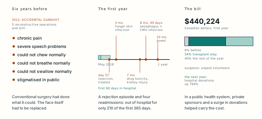 A gunshot took his face. Canada's first face transplant gave him a new one.An accidental gunshot left a man unable to chew, swallow or speak normally. In 2018 he received Canada's first face transplant, and his surgeons counted every dollar.Read it →
Want the next case like this?
When a new case is written up, it comes to your inbox: the story, the diagram and what it teaches. No schedule and no filler. Unsubscribe in one click.
Free · no spam · your address is never shared · built with KitRead the original paper: doi.org/10.12659/AOT.900617
Adapted from Krakowczyk Ł, Maciejewski A, Szymczyk C, Oleś K and Półtorak S, “Face Transplant in an Advanced Neurofibromatosis Type 1 Patient,” Annals of Transplantation 2017;22:53–57, from the Department of Oncological and Reconstructive Surgery, Maria Skłodowska-Curie Institute Cancer Center, Gliwice, Poland. The patient’s history, the donor, the operation, the immunosuppression and the recovery are as reported there, summarised in the author’s own words; the points on blood loss, vessels and nerves are those the authors make in their discussion, and the explanations of neurofibromatosis and the anatomy are from standard references. The paper is published under a Creative Commons Attribution Non-Commercial No Derivatives licence (CC BY-NC-ND 4.0), so its photographs are not reproduced here; they can be seen in the original paper. The diagram is original to MedicaseHub, is simplified, and may be reused freely under CC BY 4.0. This article is not medical advice. Read the full disclaimer.
Related cases
- A gunshot took his face. Canada's first face transplant gave him a new one.Another face transplant, and what its first year cost.
- He waited 17 months for a face that matched his ownAnother long wait for the right donor.
- The tumour covered half her face. Surgeons rebuilt it from the tumour's own skin.Another huge facial tumour, removed in a single operation.