Her whole scalp came off in one piece. Nine years later, her hair is back.
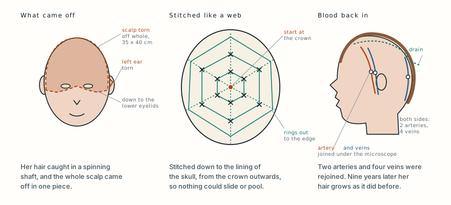
A 36-year-old woman’s hair caught in the rotating shaft of a machine, and her entire scalp was torn away, from the back of her head to her eyebrows and lower eyelids. Surgeons sewed it back on, rejoined its arteries and veins under a microscope, and pinned it to her skull in a spiderweb of stitches. Nine years later her hair grows as it did before.
- Specialty
- Microsurgery, plastic surgery
- Patient
- Woman, 36
- Mechanism
- Hair caught in the rotating shaft of a machine
- Injury
- Entire scalp torn off, about 35 × 40 cm, with the eyebrows and part of the eyelids; left ear torn
- To hospital
- About 3.5 h, in shock
- Operation
- Scalp sewn back; 2 arteries and 4 veins rejoined; spiderweb fixation to the skull
- Healed
- Day 10
- Back to normal life
- 2 months
- At 9 years
- Full hair growth, eyelashes, feeling restored, eyelids work normally
- 0 hthe machine
- 3.5 harrival
- surgeryputting it back
- day 10healed
- 9 yearsher hair
Caught by the hair
A 36-year-old woman’s hair caught in the rotating shaft of a machine, and her entire scalp tore away from her skull.
This is how almost all complete scalp avulsions happen: long hair caught in spinning agricultural or industrial machinery. The hair is stronger than the attachment of the scalp to the skull, so when the machine pulls, it is the scalp that gives way.
The scalp is built in layers: the skin, a dense layer carrying its blood vessels, a tough sheet of tendon called the galea, then a thin, loose layer, and finally the membrane on the surface of the bone. The loose layer is the weak point, and that is where her scalp separated. Everything above it came off together, with its blood vessels and its hair follicles, leaving the skull covered only by its thin membrane.
Thirty-five by forty centimetres
She reached hospital about three and a half hours after the injury, in shock. The scalp bleeds heavily, because it has one of the richest blood supplies of any skin in the body. The team controlled the bleeding with compression bandages and treated the shock.
The extent of the injury was enormous. The scalp had torn off from the back of her head forward to the bridge of her nose, through the middle of her eyebrows and down to her lower eyelids on both sides, and sideways to the roots of both ears and the cheekbones. The torn area measured about 35 by 40 centimetres. Her left ear was torn and badly scraped. The bones of her forehead, the top and back of her head and both temples lay exposed.
Once her blood pressure had stabilised, she went to theatre under general anaesthetic.
Two teams, two arteries, four veins
The surgeons split into two teams. One worked on the torn-off scalp: they cut the hair, disinfected it with iodine, trimmed its damaged edges and found and marked its blood vessels. The other worked on her head: cleaning and disinfecting the wound and finding the vessels the scalp would be connected to, the superficial temporal arteries and veins in front of each ear and the veins under the skin at the back of the head on both sides.
Before the vessels could be joined, the scalp had to be put in exactly the right place and kept there. This is the technique the paper is about. Starting from the crown of the head, the surgeons stitched the scalp down with single dissolving stitches in a pattern they describe as a spiderweb: spokes running out from the top of the head and rings joining them, working outwards towards the edges. On both temples they stitched the tissue under the skin down to the periosteum, the membrane covering the bone.
Then, under the operating microscope, they joined two arteries and four veins with stitches finer than a human hair, and blood flowed back into the scalp. The tissue under the skin was stitched down to the periosteum with more dissolving stitches, so that the scalp lay pressed flat against the skull everywhere. Drains were left under the scalp and along the incisions. They came out on the second day.
Why the stitching matters
By the tenth day after the operation, the scalp had healed completely. Living hair was growing from it, and two months after the operation she went back to her normal activities.
Joining the vessels is only half of a scalp replantation. The scalp is a large, heavy flap lying on a curved surface, and two things can ruin it even when the vessels are open. It can slide, which tears or kinks the tiny joined vessels. And blood can collect underneath it, a haematoma under the galea, which lifts the scalp away from its bed and cuts it off from the blood supply it would otherwise pick up from the tissues beneath.
The authors argue that the spiderweb pattern deals with both. Anchoring the scalp at many points stops it moving during and after the operation, which also made the vessel joins easier to do. It keeps the scalp in close contact with its bed everywhere, so that the two can grow a shared blood supply. And by pressing the layers together, it reduces oozing, so a haematoma has nowhere to form.
As it was before
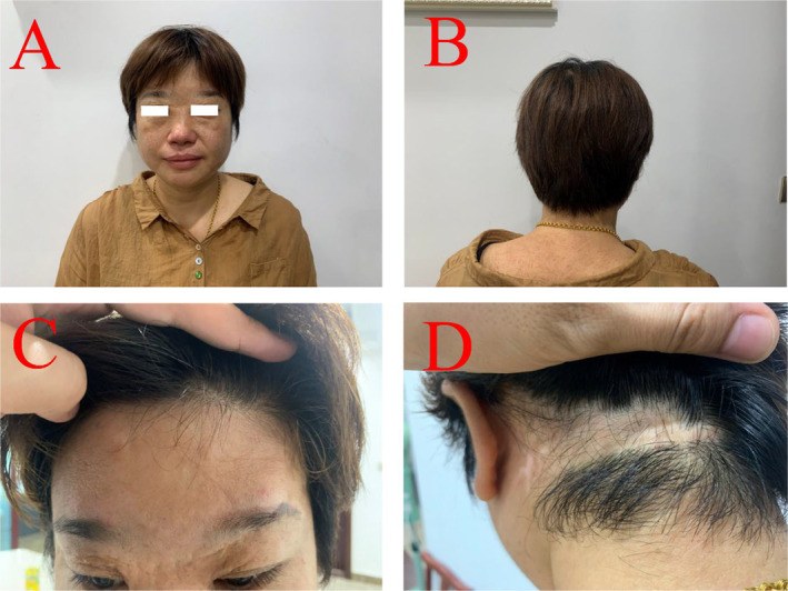
Seven months after the operation her hair was growing densely, her eyelashes were growing well and protective sensation, the ability to feel pain and pressure, had returned to her scalp. She had occasional itching of the scalp, and her right eye tended to water when she looked at something for a long time.
Nine years after the operation, the result was still good. Her hair was thick and similar to how it had been before the injury, with no patches of baldness. Her eyelashes were growing well, the scalp was soft and its protective sensation was intact, and she could open and close her eyelids without difficulty.
When the scalp cannot be saved
Before microsurgery, a torn-off scalp was usually lost. The authors list the alternatives. The scalp can be shaved down into thin skin grafts and laid back on the head, but these often fail. Where bare bone is exposed, surgeons can drill small holes in the outer skull so that a bed of healing tissue grows up through them, then graft skin onto it. Or they can move the greater omentum, a fatty apron of tissue from the abdomen, to the head with its own blood vessels and graft skin onto that. All of these can cover the skull, but they leave the head without hair.
Microsurgical replantation, reconnecting the scalp’s own vessels, is now considered the best treatment, because a scalp that lives keeps its hair follicles. The scalp is well suited to it: it is fed by several arteries running in the layer above the galea, the superficial temporal, occipital, supraorbital and supra-auricular arteries, so even a few joined vessels can keep the whole of it alive.
The technique has a clear limit, which the authors acknowledge. It only works if the torn-off scalp is in good condition, without much crushing or bruising. A scalp that has been shredded by the machine cannot be put back, however it is stitched. And the simplest prevention, for anyone who works near rotating machinery, is to keep long hair tied back and covered.
What this case teaches
A 36-year-old woman’s hair caught in a rotating machine shaft and her whole scalp, about 35 by 40 centimetres, tore away from the back of her head down to her eyebrows and lower eyelids. She arrived in shock three and a half hours later. Two surgical teams prepared the scalp and the wound, stitched the scalp back in a spiderweb pattern from the crown outwards, anchored it to the lining of the skull, and joined two arteries and four veins under a microscope. It healed in ten days, she was back to normal life in two months, and nine years later her hair, eyelashes and scalp sensation were as good as before. Reconnecting the vessels brings a scalp back to life; keeping it still and pressed flat is what lets it stay alive.
Was this case worth your time? Buy the next one a coffee ($3), or support MedicaseHub monthly.
Next case · MC-047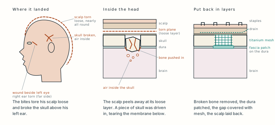 The lion was a gift. Three weeks later it attacked him.A 17-year-old was attacked by the pet lion his father had given him. He arrived in shock, with his scalp torn loose and a broken skull, and went home five days later.Read it →
Want the next case like this?
When a new case is written up, it comes to your inbox: the story, the diagram and what it teaches. No schedule and no filler. Unsubscribe in one click.
Free · no spam · your address is never shared · built with KitRead the original paper: doi.org/10.1002/ccr3.9665
Adapted from Xu L, Zhou G, Ju W, Zhang Y, Jin Q, Li L, Hou R and Ju J, “Modified Method of Scalp Fixation Promotes Survival of Complete Scalp Avulsion Replantation,” Clinical Case Reports 2024;12(12):e9665, from Suzhou Ruihua Orthopedic Hospital, Suzhou, China. The injury, the operation and its technique, and the outcome at seven months and nine years are as reported there, summarised in the author’s own words; the alternatives to replantation and the arteries of the scalp are those the authors describe in their discussion, and the explanations of the anatomy and the safety advice are from standard references. The paper is published under the Creative Commons Attribution licence (CC BY 4.0), and the authors report written consent from the patient. The photograph here is reproduced from Figure 3 under that licence, unchanged. The photographs of the injury and of the operation are not reproduced. The diagram is original to MedicaseHub, is simplified, and may be reused freely under CC BY 4.0. This article is not medical advice. Read the full disclaimer.
Related cases
- The lion was a gift. Three weeks later it attacked him.Another scalp torn loose at the same weak layer, and put back without a microscope.
- Replanting an arm: the clock, and the harder questionAnother replantation, and why the clock matters.
- They saved his ear by keeping it inside his bellyAnother torn-off part that could be saved, by a very different route.